

Duodenal Atresia
| A | Roux-en-y procedure | |
| B |
Duodenoduodenostomy |
|
| C | Duodenojejunostomy | |
| D |
duodenal canalization |
| A | Roux-en-y procedure | |
| B |
Duodenoduodenostomy |
|
| C | Duodenojejunostomy | |
| D |
duodenal canalization |
Duodenoduodenostomy REF: Bailey and love 24th ed p. 1198
The most common method of treatment of duodenal atresia is duodenoduodenostomy or duodenojejunostomy.
Double bubble sign is seen in?
| A | Duodenal atresia | |
| B |
Pyloric stenosis |
|
| C | Jejunal obstruction | |
| D | Heal obstruction |
Double bubble sign is seen in?
| A | Duodenal atresia | |
| B |
Pyloric stenosis |
|
| C | Jejunal obstruction | |
| D | Heal obstruction |
Duodenal atresia REF: Bailey and love 24T0 ed p. 1199, Nelson’s pediatrics 17th ed p. 1234
Double bubble sign is seen in
- Ladd’s bands, malrotation
- Annular pancreas
- Duodenal atresia, stenosis, web, duplication, obstruction
Anomaly associated with duodenal atresia is?
| A | Down syndrome | |
| B |
Duodenal adenomas |
|
| C | Limb defects | |
| D |
Autoimmune disorders |
Anomaly associated with duodenal atresia is?
| A | Down syndrome | |
| B |
Duodenal adenomas |
|
| C | Limb defects | |
| D |
Autoimmune disorders |
Down syndrome REF: Sabiston 18′h ed chapter 71
Duodenal atresia (DA) is thought to occur as a result of failure of vacuolization of the duodenum from its solid cord stage. The range of anatomic variants includes duodenal stenosis, mucosal web with intact muscular wall (so-called windsock deformity); two ends separated by a fibrous cord, or complete separation with a gap within the duodenum.
Associated Anomalies: DA is associated with several conditions, including prematurity, Down syndrome, maternal polyhydramnios, malrotation, annular pancreas, and biliary atresia. Other anomalies, such as cardiac, renal, esophageal, and anorectal anomalies, are also common. In most cases, the duodenal obstruction is distal to the ampulla of Vater, and infants present with bilious emesis in the neonatal period. In patients with a mucosal web, the symptoms of postprandial emesis may occur later in life.
Diagnosis: The classic plain abdominal radiograph of DA is termed the double-bubble sign (air-filled stomach and duodenal bulb, In cases in which there is no distal air, the diagnosis is secured, and no further studies are necessary. On the other hand, if distal air is present, an upper gastrointestinal contrast study is performed fairly rapidly. This study is important not only to confirm the diagnosis of duodenal stenosis or atresia but also to exclude midgut volvulus, which would constitute a surgical emergency.
Treatment: The management of DA is by surgical bypass of the duodenal obstruction as either a side-to-side or proximal transverse—to—distal longitudinal (diamond-shaped) duodenoduodenostomy.
| A | Pyloric stenosis | |
| B | Duodenal atresia | |
| C |
Ileal atresia |
|
| D | Esophageal atresia |
| A | Pyloric stenosis | |
| B | Duodenal atresia | |
| C |
Ileal atresia |
|
| D | Esophageal atresia |
Duodenal atresia [Ref: Nelson 18/e p1559; Journal- Radiology. 2001;220:463-464.]
- “The ultrasound hallmark of duodenal atresia is the ‘double bubble sign’. This consists of two adjacent. fluid-filled cavities in the upper abdomen: a lateral and usually somewhat larger cavity representing the dilated stomach, and a medial cavity representing the distended proximal duodenum between the pylorus and the stenosis.”- Ultrasound in obstetrics and gynecology By Eberhard Merz, F. Bahlmann 2/e p298
Double bubble sign:
- The double bubble sign is a finding observed on radiographs of newborns and infants. It is reproducible with a variety of other imaging modalities, including upper gastrointestinal studies and prenatal sonography. The finding depicts two air-filled structures in the upper abdomen, with little or no air distally. Horizontal-beam radiographs will demonstrate air and fluid levels in both bubbles.
- The first bubble (proximal and left sided) is – stomach
- The second bubble ( to the right of the midline) is – duodenum
- The detection of the double bubble sign leads to suspicion of a number of structural anomalies, all of which cause duodenal obstruction.
Causes:
|
Intrinsic causes are – duodenal atresia, – duodenal stenosis, – duodenal webs; |
Extrinsic causes are – annular pancreas, – malrotation of the gut with obstruction produced by midgut volvulus or by Ladd bands, and – preduodenal position of the portal vein. |
- The specific duodenal structural anomaly causing the obstruction determines the appearance, prominence, and presence of air distal to the obstruction. Duodenal atresia produces complete obstruction, Partially obstructing anomalies, such as duodenal web or duodenal stenosis, allow passage of some air distal to the obstruction and a less dramatic double bubble.
| A | Colon carcinoma | |
| B | Duodenal atresia | |
| C | Acute pancreatitis | |
| D | Perforation |
| A | Colon carcinoma | |
| B | Duodenal atresia | |
| C | Acute pancreatitis | |
| D | Perforation |
Duodenal atresia
Double-Bubble” sign in X-ray is seen in:
| A | Colon carcinoma | |
| B | Duodenal atresia | |
| C | Acute pancreatitis | |
| D | Perforation |
Double-Bubble” sign in X-ray is seen in:
| A | Colon carcinoma | |
| B | Duodenal atresia | |
| C | Acute pancreatitis | |
| D | Perforation |
Duodenal atresia
| A |
|
||||
| B |
|
||||
| C | Acute pancreatitis | ||||
| D | Perforation |
| A |
|
||||
| B |
|
||||
| C | Acute pancreatitis | ||||
| D | Perforation |
Duodenal atresia
A newborn presenting with intestinal obstruction and constipation showed, on abdominal X ray, multiple air fluid levels. The diagnosis is not likely to be:
| A |
Pyloric obstruction |
|
| B |
Duodenal atresia |
|
| C |
Ileal atresia |
|
| D |
Ladd’s bands |
A newborn presenting with intestinal obstruction and constipation showed, on abdominal X ray, multiple air fluid levels. The diagnosis is not likely to be:
| A |
Pyloric obstruction |
|
| B |
Duodenal atresia |
|
| C |
Ileal atresia |
|
| D |
Ladd’s bands |
In Pyloric obstruction there will be only single air fluid level. For multiple air fluid levels on X ray obstruction should be distal to duodenal bulb.
Ref: Walker’s Pediatric Gastrointestinal Disease: Physiology, Diagnosis and Management By Ronald E. Kleinman, 2008, Page 122
Double-bubble sign on abdominal X-ray is seen in :
| A |
Colon carcinoma |
|
| B |
Duodenal atresia |
|
| C |
Acute pancreatitis |
|
| D |
Perforation |
Double-bubble sign on abdominal X-ray is seen in :
| A |
Colon carcinoma |
|
| B |
Duodenal atresia |
|
| C |
Acute pancreatitis |
|
| D |
Perforation |
A newborn had a bilious vomiting after 6 hours of birth. X-ray of the abdomen shows double bubble sign. This is a feature of:
| A |
Pyloric stenosis |
|
| B |
Duodenal atresia |
|
| C |
Ileal atresia |
|
| D |
Esophageal atresia |
A newborn had a bilious vomiting after 6 hours of birth. X-ray of the abdomen shows double bubble sign. This is a feature of:
| A |
Pyloric stenosis |
|
| B |
Duodenal atresia |
|
| C |
Ileal atresia |
|
| D |
Esophageal atresia |
Duodenal atresia is due to failure of recanalization of the duodenal lumen during weeks 3–7 of embryologic development.
Newborn will have bilious vomiting in the first 24 hours of life.
“Double-bubble” sign and absence of gas distal to the duodenum can be seen on plain films.
A postnatal X-ray of abdomen of a neonate shows ‘double bubble sign’. It is seen with:
| A |
Pyloric stenosis |
|
| B |
Duodenal atresia |
|
| C |
Ileal atresia |
|
| D |
Esophageal atresia |
A postnatal X-ray of abdomen of a neonate shows ‘double bubble sign’. It is seen with:
| A |
Pyloric stenosis |
|
| B |
Duodenal atresia |
|
| C |
Ileal atresia |
|
| D |
Esophageal atresia |
In duodenal atresia, postnatal abdominal plain radiographs show gaseous distention of the stomach and proximal duodenum (the “double-bubble” radiologic sign).
With protracted vomiting, there is less air in the stomach and less abdominal distention.
Most common cause ofAcute Intestinal Obstruction in neonates is –
| A |
Jejuna! atresia |
|
| B |
Malrotation |
|
| C |
Duodenal atresia |
|
| D |
Acute Intussusception |
Most common cause ofAcute Intestinal Obstruction in neonates is –
| A |
Jejuna! atresia |
|
| B |
Malrotation |
|
| C |
Duodenal atresia |
|
| D |
Acute Intussusception |
Ans. is ‘c’ i.e., Duodenal atresia
o Most common cause of neonatal intestinal obstruction Meconium plug syndrome.
o Second most common cause of neonatal intestinal obstruction Duodenal atresia.
Important causes of intestinal obstruction in newborn
- Meconium plug syndrome o Duodenal atresia o Jujunoileal atresia
- Malrotation of gut o Meconium ileus
Meconium plug syndrome —> when meconium plug causes obstruction of colon. It is seen in Hirschsprung disease, maternal DM, maternal preeclampsia, prematurity, sepsis, hypothyroidism.
Meconium Ileus —> when meconium plug obstruct ileum. It is seen in cystic fibrosis.
Note ‑
o Intussusception is the most common cause of intestinal obstruction between 3 months and 6 year of age (not in neonate).
Commonest cause of intestinal obstruction in down’s syndrome-
| A |
Colomic atresia |
|
| B |
Intestinal atresia |
|
| C |
Duodenal atresia |
|
| D |
b and c |
Commonest cause of intestinal obstruction in down’s syndrome-
| A |
Colomic atresia |
|
| B |
Intestinal atresia |
|
| C |
Duodenal atresia |
|
| D |
b and c |
Ans. is ‘b i.e., Intestinal atresia & `c’ i.e., Duodenal atresia
- Most common cause of Intestinal obstruction in Down’s syndrome is duodenal atresia.
- Duodenal atresia is a type of intestinal atresia.
Double bubble sign seen in:
| A |
Duodenal atresia |
|
| B |
Ileal atresia |
|
| C |
Pyloric stenosis |
|
| D |
Pancreatic divisum |
Double bubble sign seen in:
| A |
Duodenal atresia |
|
| B |
Ileal atresia |
|
| C |
Pyloric stenosis |
|
| D |
Pancreatic divisum |
A i.e. Duodenal atresia
Double bubble sign on X-Ray is seen in:
| A |
Duodenal atresia |
|
| B |
Oesophageal atresia |
|
| C |
Colonic atresia/Hirschprung’s disease |
|
| D |
Pyloric stenosis |
Double bubble sign on X-Ray is seen in:
| A |
Duodenal atresia |
|
| B |
Oesophageal atresia |
|
| C |
Colonic atresia/Hirschprung’s disease |
|
| D |
Pyloric stenosis |
A i.e. Duodenal atresia
Antenatal double bubble appearance on ultrasound is due to –
| A |
Diaphragmatic hernia |
|
| B |
Duodenal atresia |
|
| C |
Gastric volvulus |
|
| D |
Intussusception |
Antenatal double bubble appearance on ultrasound is due to –
| A |
Diaphragmatic hernia |
|
| B |
Duodenal atresia |
|
| C |
Gastric volvulus |
|
| D |
Intussusception |
Answer is ‘b’ i.e. Duodenal atresia
- Antenatal double-bubble appearance on ultrasound is seen in duodenal obstruction, the two bubbles being the fluid filled stomach and the proximal duodenum.
Causes of duodenal obstruction in the newborn are : duodenal atresia duodenal stenosis
– duodenal web annular pancreas
– duodenal duplication cyst malrotation and midgut volvulus
- Duodenal obstruction is usually associated with polyhdyramnios.
- In 85% of infants with duodenal obstruction, the opening of bile duct is proximal to the site of obstruction. In these cases the vomiting is bilious.
- The surgical treatment of choice for duodenal atresia, stenosis or annular pancreas is duodenoduodenostomy.
Double bubble sign seen in :
| A |
Duodenal atresia |
|
| B |
Duodenal stenosis |
|
| C |
Volvulus |
|
| D |
All |
Double bubble sign seen in :
| A |
Duodenal atresia |
|
| B |
Duodenal stenosis |
|
| C |
Volvulus |
|
| D |
All |
Ans. is ‘a’, ‘b’ & ‘c’ i.e. Duodenal atresia, Duodenal stenosis and Volvulus
In case of new born the commonest cause of intestinal obstruction is –
| A |
Annular pancreas |
|
| B |
Duodenal Atresia |
|
| C |
Jejunal atresia |
|
| D |
Oesophageal atresia |
In case of new born the commonest cause of intestinal obstruction is –
| A |
Annular pancreas |
|
| B |
Duodenal Atresia |
|
| C |
Jejunal atresia |
|
| D |
Oesophageal atresia |
Ans is ‘b’ ie Duodenal atresia
- Congenital atresia and stenosis are the commonest cause.
- The incidence of atresia varies with anatomical site:
|
• |
duodenum |
35 |
percent |
|
• |
jejunum |
15 |
percent |
|
• |
ileum |
25 |
percent |
|
• |
ascending colon |
10 |
percent |
|
• |
multiple sites |
15 |
percent |
- Vomitting is bile stained or not, depends on the atresia proximal or distal to ampulla of vater. In 85% of cases the atresia is distal to the ampulla, therefore the vomitting is bilious.
- Approximately 1/3 of newborns with duodenal atresia have associated Down’s syndrome*.
- The ‘double bubble* seen on the air contrast upper GI series is characterstic of duodenal atresia.
- Treatment of choice is – Duodenoduodenostomy*
The treatment of choice in duodenal atresia ‑
| A |
Gastrojejunostomy |
|
| B |
Duodenojejunostomy |
|
| C |
Bishop koop prodcedure |
|
| D |
Duodenoduodenostomy |
The treatment of choice in duodenal atresia ‑
| A |
Gastrojejunostomy |
|
| B |
Duodenojejunostomy |
|
| C |
Bishop koop prodcedure |
|
| D |
Duodenoduodenostomy |
Ans. is ‘d’ i.e., Duodeno duodenostomy
Double Bubble sign is seen with:
| A |
Pyloric stenosis |
|
| B |
Duodenal atresia |
|
| C |
Ileal atresia |
|
| D |
Esophageal atresia |
Double Bubble sign is seen with:
| A |
Pyloric stenosis |
|
| B |
Duodenal atresia |
|
| C |
Ileal atresia |
|
| D |
Esophageal atresia |
Ans is b i.e. Duodenal atresia
“The ultrasound hallmark of duodenal atresia is the ‘double bubble sign’. This consists of two adjacent, fluid-filled cavities in the upper abdomen: a lateral and usually somewhat larger cavity representing the dilated stomach, and a medial cavity representing the distended proximal duodenum between the pylorus and the stenosis.”
Double bubble sign:
- The double bubble sign is a finding observed on radiographs of newborns and infants. It is reproducible with a variety of other imaging modalities, including upper gastrointestinal studies and prenatal sonography. The finding depicts two air-filled structures in the upper abdomen, with little or no air distally. Horizontal-beam radiographs will demonstrate air and fluid levels in both bubbles.
- The first bubble (proximal and left sided) is – stomach
- The second bubble ( to the right of the midline) is – duodenum
- The detection of the double bubble sign leads to suspicion of a number of structural anomalies, all of which cause duodenal obstruction.
Causes:
|
Intrinsic causes are |
Extrinsic causes are |
|
– duodenal atresia, |
– annular pancreas, |
|
– duodenal stenosis, |
– malrotation of the gut with obstruction |
|
and |
produced by |
|
– duodenal webs; |
midgut volvulus or by Ladd bands, and |
|
|
– preduodenal position of the portal vein. |
- The specific duodenal structural anomaly causing the obstruction determines the appearance, prominence, and presence of air distal to the obstruction. Duodenal atresia produces complete obstruction, Partially obstructing anomalies, such as duodenal web or duodenal stenosis, allow passage of some air distal to the obstruction and a less dramatic double bubble.
Double Bubble sign on abdominal skiagram is suggestive of:
March 2005
| A |
Ileal atresia |
|
| B |
Anorectal malformation |
|
| C |
Bladder calculi |
|
| D |
Duodenal Atresia |
Double Bubble sign on abdominal skiagram is suggestive of:
March 2005
| A |
Ileal atresia |
|
| B |
Anorectal malformation |
|
| C |
Bladder calculi |
|
| D |
Duodenal Atresia |
Ans. D: Duodenal Atresia
Plain films are useful in assessing the level of the obstruction.
Air-fluid levels in the stomach and duodenum (double-bubble sign), an important finding of acute duodenal obstruction, can also be seen on plain films.
Double bubble sign is a radiological feature of:
| A |
Pyloric stenosis |
|
| B |
Duodenal atresia |
|
| C |
Hirschsprung’s disease |
|
| D |
Necrotising enterocolitis |
Double bubble sign is a radiological feature of:
| A |
Pyloric stenosis |
|
| B |
Duodenal atresia |
|
| C |
Hirschsprung’s disease |
|
| D |
Necrotising enterocolitis |
Ans. Duodenal atresia
“Double bubble” sign with air shadows absent in distal bowel coils on X-ray abdomen is characteristic of
| A |
Duodenal webs |
|
| B |
Duodenal atresia |
|
| C |
CHPS |
|
| D |
All of the above |
“Double bubble” sign with air shadows absent in distal bowel coils on X-ray abdomen is characteristic of
| A |
Duodenal webs |
|
| B |
Duodenal atresia |
|
| C |
CHPS |
|
| D |
All of the above |
Ans. Duodenal atresia
In which condition,the following type of X ray is seen?

| A |
Duodenal webs |
|
| B |
Duodenal atresia |
|
| C |
Duodenal Stenosis |
|
| D |
All of the Above |
In which condition,the following type of X ray is seen?
| A |
Duodenal webs |
|
| B |
Duodenal atresia |
|
| C |
Duodenal Stenosis |
|
| D |
All of the Above |
Ans:D.)All of the above.
The Image Shows Double Bubble Sign.
The double bubble sign
It is seen in infants and represents dilatation of the proximal duodenum and stomach. It is seen in both radiographs and ultrasound, and can be identified antenatally .
Causes include
- congenital obstruction
- duodenal web
- duodenal atresia
- duodenal stenosis
- annular pancreas
- midgut volvulus
- external compression of the dudenum
- choledochal cyst
- mesenteric duplication cyst
- intramural duodenal haematoma
- preduodenal portal vein
- retroperitoneal tumour
- superior mesenteric artery syndrome
A newborn had a bilious vomiting after 6 hours of birth. X-ray of the abdomen shows as represented in the picture below. This is a feature of ? 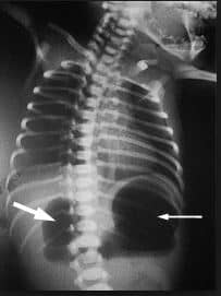
| A |
Pyloric stenosis. |
|
| B |
Duodenal atresia. |
|
| C |
Ileal atresia. |
|
| D |
Esophageal atresia. |
A newborn had a bilious vomiting after 6 hours of birth. X-ray of the abdomen shows as represented in the picture below. This is a feature of ?
| A |
Pyloric stenosis. |
|
| B |
Duodenal atresia. |
|
| C |
Ileal atresia. |
|
| D |
Esophageal atresia. |
Ans:B.)Duodenal atresia
“Double bubble” sign is seen in the image .
-Thin arrow: dialated stomach
-Thick arrow: dialated proximal duodenum
Double bubble sign
- It is seen in infants and represents dilatation of the proximal duodenum and stomach.
- It is seen in both radiographs and ultrasound, and can be identified antenatally .
- Causes include :
- congenital obstruction
- duodenal web
- duodenal atresia
- duodenal stenosis
- annular pancreas
- midgut volvulus
- external compression of the dudenum
- choledochal cyst
- mesenteric duplication cyst
- intramural duodenal haematoma
- preduodenal portal vein
- retroperitoneal tumour
- congenital obstruction
Duodenal Atresia
- It is thought to represent a failure of recanalisation of the bowel lumen that is a solid tube early in fetal life (~11-12 weeks).
- Approximately 20–40 percent of all infants with duodenal atresia have Down syndrome.
- During pregnancy, duodenal atresia is associated with increased amniotic fluid in the uterus, which is called polyhydramnios.
- After birth, duodenal atresia may cause abdominal distension, especially of the upper abdomen.
- Bilious vomiting commonly occurs within the first day of life.
- The diagnosis of duodenal atresia is usually confirmed by radiography.
- An X-ray of the abdomen shows two large air filled spaces, the so-called “double bubble” sign. The air is trapped in the stomach and proximal duodenum, which are separated by the pyloric sphincter, creating the appearance of two bubbles visible on x-ray.
- Treatment
- Early treatment includes removing fluids from the stomach via a nasogastric tube, and providing fluids intravenously. The definitive treatment for duodenal atresia is surgery (duodenoduodenostomy), which may be performed openly or laparoscopically.

