
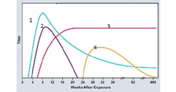
Hepatitis B Acute & Chronic Infections
All of the following are seen in active chronic hepatitis B except,
| A |
IgM against core antigen |
|
| B |
Total core antibody |
|
| C |
HbeAg |
|
| D |
HbsAg |
All of the following are seen in active chronic hepatitis B except,
| A |
IgM against core antigen |
|
| B |
Total core antibody |
|
| C |
HbeAg |
|
| D |
HbsAg |
IgM against core antigen [Ref Harrison 16th/e p. 1832]
- What is chronic active hepatitis ? . Chronic hepatitis B is usually classified by histological features such as grade and stage.
- The other way to classify chronic hepatitis is based on relative level of HBV replication :A) A Replicative phase of chronic hepatitis B is known as chronic active hepatitisQ.
. Characterized by the presence of the markers of HBV replication such as HBeAg and HBVDNA.
B) Non replicative phase of chronic hepatitis
- This is characterized by the absence of conventional markers of HBV replication i.e., (HBeAg and HBVDNA).
Thus the serological markers of chronic active hepatitis B are 😕
- HBsAg
- HBeAg
- HBVDNA
- IgGHBcAg
“IgM AntiHBcAg is nevere seen in chronic hepatits, its presence confirms that hepatitis is acute”.
Antibodies to HBcAg
Antibodies to HBcAg are not protective but we can use them to understand how long the infection has been going.
- IgM antiHBcAg Acute infection
- 1gM antiHBcAg = Chronic infection
The role of IgM antillBc in the diagnosis
- The presence of HBsAg with or without 1gM anti HBc represents HBV infection.e
- If IgM anti HBc is present, the HBV infection is considered acute and if IgM antiHBc is absent the infection is considered chronic.e
- A diagnosis of acute hepatitis can be made even in the absence of HBsAg if IgM Anti HBc is detectable.

The following is a marker of acute hepatitis B infection
| A |
DNA polymerase |
|
| B |
Hepatitis core antigen |
|
| C |
Anti HBs |
|
| D |
IgG to core antigen |
The following is a marker of acute hepatitis B infection
| A |
DNA polymerase |
|
| B |
Hepatitis core antigen |
|
| C |
Anti HBs |
|
| D |
IgG to core antigen |
DNA polymerase [Ref: Harrison 161h/e p. 1933, 1934; 16th/e p. 1832]
“DNA polymerase activity, HBVDNA, HBeAg which are representative of the vireinic stage of hepatitis B occur early in the incubation period concurrently or shortly after the first appearance of HBsAg” –
Jawetz – 19% p. 475.
- DNA polymerase is a marker for active viral replication.
- It is not exclusively associated with acute hepatitis B.
- It is also seen in chronic hepatitis B when there is active viral replication in chronic hepatitis (Chronic active hepatitis).
- But since active viral replication is usually associated with acute hepatitis, DNA polymerase is usually associated with seen in acute heptitis B.
Serological markers of Acute Hepatitis B infection
- HBsAg appears before the onset of symptomse, peaks during overt disease and then declines to undetectable levels in 3-6 months.
HBeAg, HBV-DNA and DNA polymerase appear in the serum soon after HBsAg and all signify active viral replication.
- IgM anti-HBc becomes detectable in serum shortly before the onset of symptoms, concurrent with the onset of elevation of serum aminotransferases. Over months the 1gM antibody is replaced by IgG Anti HBc (wizen infection becomes chronic)
- Anti HBS is detectable shortly after the disappearance of HBeAg implying that the acute infection has peaked and the disease is on the wane.
Hepatitis core antigen never appears in the serum.
The liver biopsy in acute hepatitis due to hepatitis B virus is likely to show all of the following histopathologic changes, EXCEPT:
| A |
Ballooning change of hepatocytes |
|
| B |
Ground glass hepatocytes |
|
| C |
Focal or spotty necrosis |
|
| D |
Acidophil bodies |
The liver biopsy in acute hepatitis due to hepatitis B virus is likely to show all of the following histopathologic changes, EXCEPT:
| A |
Ballooning change of hepatocytes |
|
| B |
Ground glass hepatocytes |
|
| C |
Focal or spotty necrosis |
|
| D |
Acidophil bodies |
After passing his physical exam, a 19 year old army recruit gives urine and blood samples for further testing. Serum analysis yields elevated ALT, HBsAg, Anti-HBc, HBeAg, and bilirubin. All other values are normal. Which of the following is the hepatitis B status of this recruit?
| A |
Asymptomatic carrier |
|
| B |
Chronic active carrier |
|
| C |
Fulminant hepatitis B |
|
| D |
Recovered from acute self-limited HBV |
After passing his physical exam, a 19 year old army recruit gives urine and blood samples for further testing. Serum analysis yields elevated ALT, HBsAg, Anti-HBc, HBeAg, and bilirubin. All other values are normal. Which of the following is the hepatitis B status of this recruit?
| A |
Asymptomatic carrier |
|
| B |
Chronic active carrier |
|
| C |
Fulminant hepatitis B |
|
| D |
Recovered from acute self-limited HBV |
A patient presented with features of acute hepatitis. He had a blood transfusion few weeks ago following an accident. Which of the following is the marker of the acute phase of hepatitis B infection?
| A |
Anti HBc (+), HBsAg (+) |
|
| B |
Anti HBe (+), HBsAg (+) |
|
| C |
Anti HBS (+), HBSAg (-) |
|
| D |
HBeAg (-), HBsAg(-) |
A patient presented with features of acute hepatitis. He had a blood transfusion few weeks ago following an accident. Which of the following is the marker of the acute phase of hepatitis B infection?
| A |
Anti HBc (+), HBsAg (+) |
|
| B |
Anti HBe (+), HBsAg (+) |
|
| C |
Anti HBS (+), HBSAg (-) |
|
| D |
HBeAg (-), HBsAg(-) |
The incidence of chronic carrier state in liver disease is due to infection with the following:
| A |
HBV |
|
| B |
HAV |
|
| C |
HEV |
|
| D |
HCV |
The incidence of chronic carrier state in liver disease is due to infection with the following:
| A |
HBV |
|
| B |
HAV |
|
| C |
HEV |
|
| D |
HCV |
All are documented patterns of glomerular injury in HBV related chronic liver disease, EXCEPT:
| A |
Minimal change nephropathy |
|
| B |
Mesangial proliferative glomerulonephritis |
|
| C |
Membranoproliferative glomerulonephritis |
|
| D |
C3 glomerulopathy |
All are documented patterns of glomerular injury in HBV related chronic liver disease, EXCEPT:
| A |
Minimal change nephropathy |
|
| B |
Mesangial proliferative glomerulonephritis |
|
| C |
Membranoproliferative glomerulonephritis |
|
| D |
C3 glomerulopathy |
The association between chronic HBV infection and glomerular diseases was first described in 1971.
- Membranous nephropathy,
- Minimal change nephropathy,
- Mesangial proliferative glomerulonephritis,
- Membranoproliferative glomerulonephritis
- IgA nephropathy
Most widely accepted mechanism associated with nephropathy in chronic HBV patients is:
| A |
Cytopathic collisions induced by cellular virus infection |
|
| B |
Deposition of immune complex particles attributed to viral antigens and host antibodies |
|
| C |
Virus-induced specific immunological mechanisms damaging the kidney |
|
| D |
Indirect adverse effects of virus-induced cytokines or mediators in renal tissue. |
Most widely accepted mechanism associated with nephropathy in chronic HBV patients is:
| A |
Cytopathic collisions induced by cellular virus infection |
|
| B |
Deposition of immune complex particles attributed to viral antigens and host antibodies |
|
| C |
Virus-induced specific immunological mechanisms damaging the kidney |
|
| D |
Indirect adverse effects of virus-induced cytokines or mediators in renal tissue. |
HBV can cause renal damage in many ways. The different pathogenesis are described as the cause for it. They are,
Hera lal a 35 year old man was found to be positive for HBsAg and HBeAg , accidentally during a screening for blood donation. On Lab examination SGOT and SGPT are normal. What should be the most appropriate next step?
| A |
Observation |
|
| B |
Liver biopsy |
|
| C |
Interferon therapy |
|
| D |
HBV-DNA estimation |
Hera lal a 35 year old man was found to be positive for HBsAg and HBeAg , accidentally during a screening for blood donation. On Lab examination SGOT and SGPT are normal. What should be the most appropriate next step?
| A |
Observation |
|
| B |
Liver biopsy |
|
| C |
Interferon therapy |
|
| D |
HBV-DNA estimation |
Patient in the question is positive for HBsAg and HBeAg, so he is more likely to be highly infectious and must be associated with the presence of Hepatitis B virions and a detectable HBV DNA.
Ref: Harrisons Internal Medicine, 18th Edition, Chapter 304, Page 2540 ; Review of Medical Microbiology and Immunology By Warren Levinson, 11th Edition, Chapter 41
A male patient is observed to be HBs Ag antigen positive HBe Ag antigen negative and anti-HBe antibody positive. HBV DNA copies are observed to be 100,000/ml while SGOT and SGPT are elevated to 6 times the upper limit of normal value. What is the likely diagnosis?
| A |
HBV surface mutant |
|
| B |
HBV precore mutant |
|
| C |
Wild HBs Ag |
|
| D |
Inactive HBV carrier |
A male patient is observed to be HBs Ag antigen positive HBe Ag antigen negative and anti-HBe antibody positive. HBV DNA copies are observed to be 100,000/ml while SGOT and SGPT are elevated to 6 times the upper limit of normal value. What is the likely diagnosis?
| A |
HBV surface mutant |
|
| B |
HBV precore mutant |
|
| C |
Wild HBs Ag |
|
| D |
Inactive HBV carrier |
Patient in the question stem is showing features of chronic active hepatitis. HBeAg is negative due to the pre core mutant variant of hepatitis B. Pre core mutation is responsible for the decrease or non production of HBeAg. Such mutations often occur at the time of seroconversion from HBe Ag to anti HBe Ab.
Ref: Digestive Involvement in Systemic Autoimmune Diseasesedited by Josep Font, Page 192; Detection of Bacteria, Viruses, Parasites and Fungi: Bioterrorism Prevention By Mariapia Viola Magni, Page 248; Sherlock’s Diseases of the Liver and Biliary System edited by James S. DooleypGE 367, 8.
About chronic hepatitis B , all are true, EXCEPT:
| A |
90 % chance for chronic infection if infected at birth. |
|
| B |
Mild chronic hepatitis B has 5 year survival 97 % |
|
| C |
Adult infection is usually symptomatic |
|
| D |
Chronicity if infected in adult age is about 10 % |
About chronic hepatitis B , all are true, EXCEPT:
| A |
90 % chance for chronic infection if infected at birth. |
|
| B |
Mild chronic hepatitis B has 5 year survival 97 % |
|
| C |
Adult infection is usually symptomatic |
|
| D |
Chronicity if infected in adult age is about 10 % |
Chronic hepatitis B:
HBV is associated with all of the following except ‑
| A |
Hepatic cancer |
|
| B |
Chronic hepatitis |
|
| C |
Hepatic adenoma |
|
| D |
Cirrhosis |
HBV is associated with all of the following except ‑
| A |
Hepatic cancer |
|
| B |
Chronic hepatitis |
|
| C |
Hepatic adenoma |
|
| D |
Cirrhosis |
Ans. is ‘c’ i.e., Hepatic adenoma
Complications of Acute HBV infection
. Fulminant hepatitis . Cirrhosis
. Hepatocellular carcinoma . Chronic hepatitis
. Papular acrodermatitis of child hood (Gianotti – crosti syndrome) . Glomerulonephritis
. Essential mixed cryoglobulinemia (more common with HCV)
. Polyarteritis nodosa (PAN) may be associated with HBV infection.
Remember
Hepatocellular carcinoma is the only vaccine preventable cancer.
A 30 year old patient presented with history of jaundice for 10 days. His liver function tests showed bilirubin of 10 mg/dl, SGOT/SGPT – 1100/1450, serum alkaline phosphatase – 240 LU. He was positive for Hbs Ag. What should be the confirmatory test to estabilish acute hepatitis B infection –
| A |
Ig M anti-HBc antibody |
|
| B |
Hbe Ag |
|
| C |
HBV DNV by PCR |
|
| D |
Anti-HBc antibody |
A 30 year old patient presented with history of jaundice for 10 days. His liver function tests showed bilirubin of 10 mg/dl, SGOT/SGPT – 1100/1450, serum alkaline phosphatase – 240 LU. He was positive for Hbs Ag. What should be the confirmatory test to estabilish acute hepatitis B infection –
| A |
Ig M anti-HBc antibody |
|
| B |
Hbe Ag |
|
| C |
HBV DNV by PCR |
|
| D |
Anti-HBc antibody |
Ans. is ‘a’ i.e., Ig M anti-HBc antibody
. Has been explained
. Ig M anti-HBc antibody is the marker of acute or recent HBV infection.
Best means of giving hepatitis B vaccine is
| A |
Subcutaneous |
|
| B |
Intradermal |
|
| C |
Intramuscular deltoid |
|
| D |
Intramuscular gluteal |
Best means of giving hepatitis B vaccine is
| A |
Subcutaneous |
|
| B |
Intradermal |
|
| C |
Intramuscular deltoid |
|
| D |
Intramuscular gluteal |
Ans. is ‘c’ i.e., Intramuscular Deltoid
Prophalyxis against HBV
. Both passive and active immunization are available for HBV infection prophylaxis.
. Passive prophylaxis is by hepatitis B immune globulin (HBIG).
. Active immunization is by recombinant hepatitis B vaccine.
. These are two recombinant vaccines :
- Recombivax – HB
- Engerix – B
- Vaccine is given intramuscular into the deltoid or in infants into the anterolateral aspect of thigh. Gluteal injection is not recommended as it may result in poor immune response.
. Three doses are given at 0, 1 and 6 months.
. For pre exposure prophylaxis only hepatitis B vaccine is given.
. For post exposure prophylaxis combination of HBIG and hepatitis B vaccine is recommended.
- For perinatal exposure single dose of HBIG at birth along with complete course of vaccination is recommended. First dose of vaccine should be given within 12 hours after birth.
The liver biopsy in acute hepatitis due to hepatitis B virus is likely to show all of the following, except ‑
| A |
Ballooning change of hepatocytes |
|
| B |
Ground glass hepatocytes |
|
| C |
Focal or spotty necrosis |
|
| D |
Acidophil bodies |
The liver biopsy in acute hepatitis due to hepatitis B virus is likely to show all of the following, except ‑
| A |
Ballooning change of hepatocytes |
|
| B |
Ground glass hepatocytes |
|
| C |
Focal or spotty necrosis |
|
| D |
Acidophil bodies |
Ans. is ‘b’ i.e., Ground glass hepatocytes
- Ground glass hepatocytes are large hepatocytes containing surface antigen. Their cytoplasm is ground glass in appearance
o These cells are seen in chronic hepatitis (not acute hepatitis)
o Ground glass hepatocytes can be identified histochemically with orcein or aldehyde fuschin stain.
Pathological features of acute viral hepatitis
1. Panlobular infiltration of mononuclear cells
This mononuclear infiltration primarily consists of —> Lymphocytes, Plasma cells, Eosinophils
Inflammatory infiltrate may spill over into the adjacent parenchyma to cause necrosis of periportal hepatocytes -3 interface hepatitis, can occur in both acute and chronic hepatitis.
2. Hepatic cell damage – It consits of
(a) Hepatic cell necrosis – The necrosis is usually focal or centirzonal.
Sometimes severe necrosis such as bridging or subacute hepatic necorsis occurs.
(b) Ballooning of cells — Damaged cells show diffuse swelling known as ballooning degeneration.
(c) Acidophilic degeneration of hepatocytes – In this single necrotic liver cell has coagulated pink cytoplasm and it shows pyknosis or karyolysis. These are called councilman bodies.
(d) Cell dropout
3. Hyperplasia of kupffer cells
4. Variable degree of cholestasis with brown pigmentation of hepatocytes.
5. Hepatic cell regeneration – It is indicated by increased mitotic figures, rosette or pseudoacinar formation and multi nucleated cells.
6. Lobular disarray —> Loss of normal lobular architecture.
o Note that these morphological lesions are similar in all types of viral hepatitis.
o A few histological changes may be indicative of particular type of virus.
1.In hepatitis C the histological lesion is characterized by –
a Relative paucity of inflammation
Marked increase in activation of sinusoidal lining cells.
Presence of lymphoid aggregates
Presence offal Macrovesicular steatosis
Bile duct lesions.
2.In Hepatitis D -* Microvesicular steatosis occurs.
3.In Hepatitis E —)Marked cholestasis occurs.
Ground glass hepatocytes are seen in which of the viral hepatitis –
| A |
HAV |
|
| B |
HBV |
|
| C |
HCV |
|
| D |
HDV |
Ground glass hepatocytes are seen in which of the viral hepatitis –
| A |
HAV |
|
| B |
HBV |
|
| C |
HCV |
|
| D |
HDV |
Ans. is ‘b’ i.e., HBV
o HBV-infected hepatocytes may show a cytoplasm packed with spheres and tubules of HBs Ag, producing a finely granular cytoplasm (“ground-glass hepatocytes”).
HBV is not associated with –
| A |
Chronic active hepatitis |
|
| B |
Chronic persistent hepatitis |
|
| C |
Post necrotic cirrhosis |
|
| D |
Cholangio carcinoma |
HBV is not associated with –
| A |
Chronic active hepatitis |
|
| B |
Chronic persistent hepatitis |
|
| C |
Post necrotic cirrhosis |
|
| D |
Cholangio carcinoma |
Ans. is ‘d’ i.e., Cholangiocarcinoma
A 45 day – old infant developed icterus and two days later symptoms and signs of acute liver failure appeared. Child was found to be positive for Hbs Ag. The mother was also HBs Ag carrier. The mother’s hepatitis B serological profile is likely to be ‑
| A |
HBs Ag positive only |
|
| B |
HBs Ag and Hbe Ag positivity |
|
| C |
HBsAg and anti – HBe antibody positivity |
|
| D |
Mother infected with mutant HBV |
A 45 day – old infant developed icterus and two days later symptoms and signs of acute liver failure appeared. Child was found to be positive for Hbs Ag. The mother was also HBs Ag carrier. The mother’s hepatitis B serological profile is likely to be ‑
| A |
HBs Ag positive only |
|
| B |
HBs Ag and Hbe Ag positivity |
|
| C |
HBsAg and anti – HBe antibody positivity |
|
| D |
Mother infected with mutant HBV |
Ans. is `b’ i.e., HBs positive and Hbe Ag positive
Perinatal transmission of HBV
o Most important risk factor that determines perinatal transmission is HBe Ag. HBs Ag carrier mothers who are HBe Ag positive almost invariably (> 90%) transmit hepatitis B infection to their offspring, where as carrier mothers with anti HBe rarely (10 to 15%) infect their offspring.
o Most common time of perinatal transmission is at the time of delivery.
o Most of the infected neonates are asymptomatic carrier with increased chances of chronic hepatitis and hepatocellular carcinoma.
A 30-year old lady delivered a healthy baby at 37 week of gestation. She was a known case of chronic hepatitis B infection She was positive for HBsAG but negative for HBeAG. Which of the following is the most appropriate treatment for the baby ‑
| A |
Both active and passive immunization soon after birth |
|
| B |
Passive immunization soon afte birth and active immunization at I year of age |
|
| C |
Only passive immunization soon after birth |
|
| D |
Only active immunization soon after birth |
A 30-year old lady delivered a healthy baby at 37 week of gestation. She was a known case of chronic hepatitis B infection She was positive for HBsAG but negative for HBeAG. Which of the following is the most appropriate treatment for the baby ‑
| A |
Both active and passive immunization soon after birth |
|
| B |
Passive immunization soon afte birth and active immunization at I year of age |
|
| C |
Only passive immunization soon after birth |
|
| D |
Only active immunization soon after birth |
Ans. is ‘a’ i.e., Both active and passive immunization soon after birth
Immunization of infants born to HbsAg positive women.
o These infants should receive both active and passive immunization soon after birth.
The first dose of active immunization should be accompanied by administration of .5 nil of HBIG as
soon after delivery is possible, because the effectiveness decreases rapidly with increased time after birth.
o Subsequent doses of active immunization should be given at 1-2mo, and 6 mo of age.
True about Hepatocellular Carcinoma ‑
| A |
Most prevalent malignancy |
|
| B |
Commonest liver tumor |
|
| C |
HBV predisposes |
|
| D |
Patient present in early stage |
True about Hepatocellular Carcinoma ‑
| A |
Most prevalent malignancy |
|
| B |
Commonest liver tumor |
|
| C |
HBV predisposes |
|
| D |
Patient present in early stage |
Ans. is ‘c’ i.e. HBV predisposes
- Most common cancers in the world are —> Lung > Breast > Colorectal.
- As already stated before, HCC presents quite late and thus only 15-20% cancers are resectable. Most patients with unresectable lesions die within a year after diagnosis.
Which of the following viral infections is associated with highest risk of progression to fulminant disease and acute liver failure:
| A |
HEV infection |
|
| B |
HBV Infection |
|
| C |
HBV/HDV coinfection |
|
| D |
HDV /HBV Superinfection |
Which of the following viral infections is associated with highest risk of progression to fulminant disease and acute liver failure:
| A |
HEV infection |
|
| B |
HBV Infection |
|
| C |
HBV/HDV coinfection |
|
| D |
HDV /HBV Superinfection |
Answer is D (HDV/HBV Superinfection)
HDV superinfection of chronic Hepatitis B is associated with a 20 percent risk of progression to fulminant hepatitis and Acute Hepatic Failure.
|
Feature |
HAV |
HCV |
HEV |
HBV |
|
Fulminant Infection |
0.1% |
0.1% |
1-2 % |
HBV : 1 % |
|
(Acute Liver Failure) |
|
|
in HEV pregnant p |
HBV/HDV Co-infection : 5 % |
|
|
|
|
Women: 20% |
HDV/HBV Superinfection: 20 % |
Tendency to cause acute liver failure (fulminant infection) is highest with HDV Superinfection on Chronic Hepatitis B and Hepatitis E infection in pregnant women (up to 20 percent)
Note:
- Hepatitis E is the most common cause of Acute Liver Failure in India
- Hepatitis B remains the most common viral agent responsible for Acute Liver Failure Worldwide Although Hepatitis E has a higher tendency to cause fulminant infection (1-2 %) in comparison to Hepatitis B infection (Up-to 1%), Hepatitis B is the most common cause of fulminant infection overall since the overall incidence of Hepatitis B Hepatitis is higher than the incidence of Hepatitis E Hepatitis
- Tendency to cause acute liver failure (fidminant infection) is highest with HDV Superinfection on Chronic Hepatitis B and Hepatitis E infection in Pregnant women (up to 20 percent)
A male patient is observed to be HBs Ag antigen positive HBe Ag antigen negative and anti-HBe antibody positive. HBV DNA copies are observed to be 100,000/m1 while SGOT and SGPT are elevated to 6 times the upper limit of normal value. What is the likely diagnosis:
| A |
HBV surface mutant |
|
| B |
HBV precore mutant |
|
| C |
Wild HBs Ag |
|
| D |
Inactive HBV carrier |
A male patient is observed to be HBs Ag antigen positive HBe Ag antigen negative and anti-HBe antibody positive. HBV DNA copies are observed to be 100,000/m1 while SGOT and SGPT are elevated to 6 times the upper limit of normal value. What is the likely diagnosis:
| A |
HBV surface mutant |
|
| B |
HBV precore mutant |
|
| C |
Wild HBs Ag |
|
| D |
Inactive HBV carrier |
Answer is B (HBV pre-core mutant)
Evidence of chronic active hepatitis B (suggested by elevated liver enzymes) in a patients with detectable HBV DNA but negative HBe Ag suggests a diagnosis of precore mutant chronic hepatitis B
Precore mutant chronic hepatitis B
- Precore mutant form of chronic hepatitis B represents an infection with a molecular variant of the hepatitis B virus that results in an unusual serologic — clinical profile.
- Such patients have severe chronic hepatitis B infection with detectable HBV DNA levels, but HBe Ag antigen is typically negative
- This results from an infection with an HBV mutant that contains an alteration in the precore regions rendering the virus incapable of encoding HBe Ag
- Patients with such mutations in the precore region are unable to secrete HBc Ag, have a negative HBe Ag phenotype but tend to have severe liver disease that progresses more rapidly to cirrhosis
- Characteristic’ of such HBe Ag negative (precore mutant) chronic hepatitis B are lower levels of HBV DNA (< 105 copies /m1) and one of several patterns of aminotransferase activity — persistent elevation, periodic fluctuation above the normal range and periodic fluctuations between the normal and elevated range
- HBe Ag negative chronic hepatitis B with mutation in the pre-core region is now the most frequently encountered form of hepatitis B in the Mediterranean countries and in Europe.
|
|
HBs Ag |
Anti HBs Antibody |
Anti HBc Antibody |
HBe Ag |
Anti HBe Antibody |
HBV DNA |
|
Acute Hepatitis B (early) |
+ |
|
|
+ |
– |
+ |
|
Acute Hepatitis B – (window period) |
|
– |
IgM |
+ |
– |
+ – |
|
Acute Hepatitis B (recovery) |
+ |
+ |
IgG |
– |
+ |
‑ |
|
Chronic Hepatitis B, high replication |
|
– |
IgG |
+ |
– |
+ |
|
Chronic Hepatitis B, low/non-replicati Ye phase |
|
– |
IgG |
– |
+ |
– |
|
Chronic Hepatitis B Precore Mutant |
|
– |
IgG |
|
4- |
+ |
|
Chronic Hepatitis B inactive carrier |
+ |
– |
IgG |
– |
+7- |
Low-level + |
|
Chronic hepatitis B reactivation |
+ |
– |
IgM |
+ |
– |
|
|
Hepatitis B vaccine |
|
|
– |
– |
|
– |
A patient is found to be positive for HBs Ag on routine laboratory evaluation. Other serological tests for hepatitis are unremarkable. He is clinically asymptomatic and liver enzymes are within the normal range. Which of the following best describes his diagnosis:
| A |
Inactive HBV carrier |
|
| B |
Acute Hepatitis B |
|
| C |
Chronic Hepatitis B |
|
| D |
Active HBV carrier |
A patient is found to be positive for HBs Ag on routine laboratory evaluation. Other serological tests for hepatitis are unremarkable. He is clinically asymptomatic and liver enzymes are within the normal range. Which of the following best describes his diagnosis:
| A |
Inactive HBV carrier |
|
| B |
Acute Hepatitis B |
|
| C |
Chronic Hepatitis B |
|
| D |
Active HBV carrier |
Answer is A (Inactive HBV carrier)
Absence of clinical signs and symptoms of liver damage and normal liver enzymes in an HBs Ag positive individual with otherwise unremarkable serological tests suggests a diagnosis of Inactive HBV carrier state.
‘An inactive hepatitis B carrier is defined as one with HBs Ag in serum hut without HBe Ag with normal liver function tests and with low/ undetectable levels of HBV DNA (< 103-104 virions/ml) – ‘Blue Prints Pocket Gastroenterology’ by Grover (Lippincott Williams) 1st (2007) /114
Chronic Hepatitis B carriers (HBs Ag + ye with IgG Anti HBc)
Chronic Hepatitis B virus carriers can be divided into two categories based on activity of the virus and evidence of liver damage
Inactive HBs Ag Carrier state
- Patients with negligible virus replication (HBV DNA levels < 103 virions/m1) and /or no clinical signs and symptoms of liver damage and normal liver enzyme levels are characterized as inactive HBV carriers Inactive HBV carriers harbor Hepatitis B virus antigen (HBs Ag), but are clinically asymptomatic, have a normal physical examination with normal liver enzymes (AST & ALT). HBe Ag is typically negative and levels of HBV DNA are less than 103-104 virions/ml
- Such patients have minimal (if any) liver inflammation or damage, and usually live a normal life. However compared to the general population these patients remain at risk for hepatocellular carcinoma
- Treatment is currently not recommended for
- Inactive HBV carrier. However regular observation is advised as there is a risk for reactivation of virus.
Chronic Hepatitis B (Active)
Patients with active virus replication ( HBV DNA levels > 103 -104 virions/ml) and /or clinical signs and symptoms of liver damage or elevated liver enzyme levels are characterized as chronic hepatitis B patients.
Depending on the presence or absence of HBe Ag two type of chronic (active) hepatitis B may be identified.
HBe Ag positive chronic hepatitis B
These HBs Ag positive, individuals are also positive for HBe Ag and show detectable HBV DNA levels. Such patients are in the replicative phase of chronic hepatitis B infection, have persistently or intermittently elevated liver enzymes and liver biopsy results typically reveals inflammation and damage
Patients with chronic (active) hepatitis B may become inactive (relatively non replicative) over time (rate of L-10 % per year) by becoming negative for HBe Ag and HBV DNA (HBV DNA < 10' - 104) virions/ml) Liver injury tend to subside in such patients and these may then be characterized as ‘inactive carriers’
HBe Ag negative chronic hepatitis B (Precore mutant)
Such HBs Ag positive individuals are negative for HBe Ag but show detectable HBV DNA levels. Such patients are also in the replicative phase of chronic hepatitis B (despite negative HBe Ag) and have persistently or intermittently elevated liver enzymes and liver biopsy results typically reveal severe inflammation and damage
Negative HBe Ag Status results from a mutant strain of hepatitis B that is characterized by the .failure of the virus to make the hepatitis B ‘e’ antigen (HBe Ag). This mutation is called the `precore’ mutation as it prevents the translation of a precore protein to prevent HBe antigen production.
This mutation does not however affect the ability of the virus to replicate
The following is a marker of acute hepatitis B infection:
| A |
DNA polymerase |
|
| B |
Hepatitis core antigen |
|
| C |
Anti HBs |
|
| D |
IgG to core antigen |
The following is a marker of acute hepatitis B infection:
| A |
DNA polymerase |
|
| B |
Hepatitis core antigen |
|
| C |
Anti HBs |
|
| D |
IgG to core antigen |
Answer is A (DNA polymerase)
DNA polymerase is a marker for active viral replication.
DNA polymerase are usually associated with Acute viral hepatitis B when there is active viral replication and hence DNA polymerase is the single best answer amongst the options provided
Note that although DNA polymerase is often present in acute Hepatitis B infection, its presence suggests active virus replication (like HbeAg, HBV DNA) and it should not be used as a laboratory marker for acute hepatitis B infection.
`During the acute episode of disease when there is active viral replication large amounts of HB.sAg and Hepatic B virus DNA can be detected in the serum as can fully developed virions and high levels of DNA polymerase and HBeAg’-Current Diagnosis and Treatment in Infections Disease/437
Hera lal 35 years old man was found +ve for HBsAg & HBeAg, accidentally during screening of blood donation. On Lab examination SGOT & SGPT are Normal. What should you do next :
| A |
Liver biopsy |
|
| B |
Interferon therapy |
|
| C |
Observation |
|
| D |
HBV-DNA estimation |
Hera lal 35 years old man was found +ve for HBsAg & HBeAg, accidentally during screening of blood donation. On Lab examination SGOT & SGPT are Normal. What should you do next :
| A |
Liver biopsy |
|
| B |
Interferon therapy |
|
| C |
Observation |
|
| D |
HBV-DNA estimation |
Answer is D (HBV-DNA estimation)
Like Hbe Ag, serum HBV DNA is an indicator of HBV replication, but tests for. HBV DNA are more sensitive and quantitative- Harrison 161h/1832
The marker HBV DNA is useful in following the course of HBV replication and thus deciding need for antiviral chemotherapy.
Interferon therapy would be started after adjudging its need as indicated by HBV DNA testing.
Liver biopsy would be too radical at this stage.
Which one of the following pairs regarding Hepatitis B is not correctly matched
| A |
Acute Viral Hepatitis B – Supportive care |
|
| B |
Acute Viral Hepatitis B – Antiviral therapy |
|
| C |
Chronic Viral Hepatitis B – Supportive care |
|
| D |
Chronic Viral Hepatitis B – Antiviral therapy |
Which one of the following pairs regarding Hepatitis B is not correctly matched
| A |
Acute Viral Hepatitis B – Supportive care |
|
| B |
Acute Viral Hepatitis B – Antiviral therapy |
|
| C |
Chronic Viral Hepatitis B – Supportive care |
|
| D |
Chronic Viral Hepatitis B – Antiviral therapy |
Answer is B (Acute Viral Hepatitis B – Antiviral therapy):
Acute Viral Hepatitis B is not an indication of Antiviral therapy. Antiviral therapy is not indicated for Acute Hepatitis B.
Acute Hepatitis B is not an approved indication for Antiviral Therapy
In the vast majority of patients Acute Hepatitis B is a self limiting disease that results in viral elimination and complete healing.
‘In Hepatitis B among previously healthy adults who present with clinically apparent hepatitis, recovery occurs in – 99%; therefore antiviral therapy is not likely to improve the rate of recovery and is not required’- Harrison ‘s 17’h/1946
‘The administration of Antiviral therapy (lamiyudine) to patients with acute hepatitis B may slow the development of protective Anti- HBs titres. Therefore antiviral treatment is not recommended in patients with uncomplicated acute viral hepatitis B’ – Clinical Hepatology (Springer, 2010) /761
Note (Harrison ‘s 17th /1946; Clinical Hepatology (2010)/761)
Antiviral therapy with nucleos(t)ide analogs might be considered for fewer than I% of patients with acute viral hepatitis B who present with a Alininant clinical course. Although at present severe/fuhninant acute hepatitis B is not an approved indication of antiviral therapy and there is no data to support such an approach most authorities would recommended institution of antiviral therapy for severe acute Hepatitis B
Extrahepatic Manifestations of Hepatitis B include all of the following Except
| A |
Aplastic Anemia |
|
| B |
Gullian Barre syndrome |
|
| C |
Pancreatitis |
|
| D |
Gall stone disease |
Extrahepatic Manifestations of Hepatitis B include all of the following Except
| A |
Aplastic Anemia |
|
| B |
Gullian Barre syndrome |
|
| C |
Pancreatitis |
|
| D |
Gall stone disease |
Answer is D (Gall stone disease)
Gallstone disease has not been mentioned in the list for extrahepatic manifestations of Hepatitis B
Extrahepatic manifestations in viral hepatitis B:
- Aplastic Anemia
- Cryoglobulinaemia
- Diabetes mellitus
- Dermatomyositis-like syndrome
- Gianotti- Crosti syndrome
- Glomerulonephritis
- Gui!lain- Barre syndrome
- Myasthenia gravis
- Pericarditis and myocarditis
- Pancreatitis
- Peripheral Neuropathy
- Polyarleritis Nodosa
- Polyarthritis
- Polymyalgia rheumatica
- Polymyositis
- Porphyria cutanea tarda
- Psychosis
- Raynaud’s syndrome
- Serum sickness like illness
- Splenomegaly and lymph node enlargement
- Skin alterations
- Thrombopenic. purpura
- Thyroiditis
- Urticaria
Interferon treatment is recommended in chronic hepatitis B in patients with:
| A |
↑ HBV DNA and Normal ALT |
|
| B |
↑ HBV DNA and ↑ ALT |
|
| C |
↑ HBV DNA and compensated cirrhosis |
|
| D |
↑ HBV DNA and decompensated cirrhosis |
Interferon treatment is recommended in chronic hepatitis B in patients with:
| A |
↑ HBV DNA and Normal ALT |
|
| B |
↑ HBV DNA and ↑ ALT |
|
| C |
↑ HBV DNA and compensated cirrhosis |
|
| D |
↑ HBV DNA and decompensated cirrhosis |
Answer is B (↑ HBV DNA and ↑ ALT)
Antiviral therapy with interferon is indicated in patients with active viral replication (is HBV DNA) and elevated ALT levels
Patient with active DNA replication (T HBV DNA) and cirrhosis (compensated or decompensated) should not be treated with Interferon therapy These patients should he treated with other oral agents and IFN
Indications for Interferon Therapy
Chronic. Hepatitis with “iHBV DNA and Elevated LFT
Hepatitis B vaccine should be given as per which schedule:
September 2005
| A |
0,1,6 days |
|
| B |
0,1,6 weeks |
|
| C |
0,1,6 months |
|
| D |
0,1,6 years |
Hepatitis B vaccine should be given as per which schedule:
September 2005
| A |
0,1,6 days |
|
| B |
0,1,6 weeks |
|
| C |
0,1,6 months |
|
| D |
0,1,6 years |
Ans. C: 0, 1, 6 months
The dose for the adult is 10-20 micrograms initially and again at 1 and 6 months.
Children under 10 years should be given half of the adult doses at the same intervals or at 6, 10 and 14 weeks as per Indian National Immunization Schedule.
For greater reliability of absorption, deltoid is the preferred for injection as gluteal injection often results in deposition of the vaccine in fat rather than in muscle.
For Children under 2 years of age, anterolateral aspect of thigh is used as vaccination site.
Vaccine which should not be frozen –
| A |
OPV |
|
| B |
Measles |
|
| C |
HBV |
|
| D |
Yellow fever |
Vaccine which should not be frozen –
| A |
OPV |
|
| B |
Measles |
|
| C |
HBV |
|
| D |
Yellow fever |
Ans. is ‘c’ i.e., HBV
OPV and measles vaccines are stored in deep freezers. (Note : Yellow fever vaccine is also freez dried, but is not used in India).
Vaccine which must be stored in the cold part but never allowed to freez.
- Typhoid
- DPT
- TT
- Hepatitis B
- DT
- BCG
- Diluents
Hepatitis B associated with ‑
| A |
Wegener’s granulomatosis |
|
| B |
Systemic lupus erythmatosus |
|
| C |
Polyarteritis nodosa |
|
| D |
Sjogren syndrome |
Hepatitis B associated with ‑
| A |
Wegener’s granulomatosis |
|
| B |
Systemic lupus erythmatosus |
|
| C |
Polyarteritis nodosa |
|
| D |
Sjogren syndrome |
Ans. is ‘c’ i.e., Polyatrteritis nodosa
The hepatitis B surface antigen is present in 25% of patients with Polyarteritis Nodosa.
Hepatitis C antibody is present in patients with essential mixed cryoglobinuria and rarely in patients with poly arteritis nodosa.
Marker of level of liver injury in cases of hepatitis B is –
| A |
HBsAg |
|
| B |
HBV DNA |
|
| C |
Anti HBeAg |
|
| D |
Anti HBc |
Marker of level of liver injury in cases of hepatitis B is –
| A |
HBsAg |
|
| B |
HBV DNA |
|
| C |
Anti HBeAg |
|
| D |
Anti HBc |
Ans. is ‘b’ i.e., HBV DNA
- In patients with chronic hepatitis B infection the level of HBV DNA correlates with the level of liver injury and risk of progression of disease.
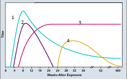
A male patient is observed to be HBs Ag antigen positive HBe Ag antigen negative and anti-HBe antibody positive. 1 marked in this grapph is observed to be 100,000/ml while SGOT and SGPT are elevated to 6 times the upper limit of normal value. What is the likely diagnosis?
| A |
HBV surface mutant |
|
| B |
HBV precore mutant |
|
| C |
Wild HBs Ag |
|
| D |
Inactive HBV carrier |
A male patient is observed to be HBs Ag antigen positive HBe Ag antigen negative and anti-HBe antibody positive. 1 marked in this grapph is observed to be 100,000/ml while SGOT and SGPT are elevated to 6 times the upper limit of normal value. What is the likely diagnosis?
| A |
HBV surface mutant |
|
| B |
HBV precore mutant |
|
| C |
Wild HBs Ag |
|
| D |
Inactive HBV carrier |
Ans is b
1 in this graph is HBV DNA
Patient in the question stem is showing features of chronic active hepatitis. HBeAg is negative due to the pre core mutant variant of hepatitis B. Pre core mutation is responsible for the decrease or non production of HBeAg. Such mutations often occur at the time of seroconversion from HBe Ag to anti HBe Ab.

