
LIVER
The right lobe of the liver consists which of the following segments?
| A |
V, VI, VII and VIII |
|
| B |
IV, V, VI, VII and VIII |
|
| C |
I, V, VI, VII and VIII |
|
| D |
I, IV, V, VI, VII and VIII |
The right lobe of the liver consists which of the following segments?
| A |
V, VI, VII and VIII |
|
| B |
IV, V, VI, VII and VIII |
|
| C |
I, V, VI, VII and VIII |
|
| D |
I, IV, V, VI, VII and VIII |
Ans. V, VI, VII and VIII
The following diagram shows the various lobes of Liver. Identify Structure Marked A in the diagram
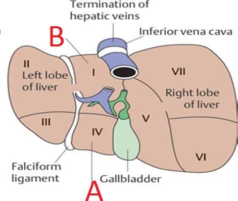
| A |
Caudate Lobe |
|
| B |
Quadrate Lobe |
|
| C |
Medial Lobe |
|
| D |
None of the Above |
The following diagram shows the various lobes of Liver. Identify Structure Marked A in the diagram
| A |
Caudate Lobe |
|
| B |
Quadrate Lobe |
|
| C |
Medial Lobe |
|
| D |
None of the Above |
Bare area of liver is related to –
| A |
Aorta |
|
| B |
Hepatic vein |
|
| C |
Portal vein |
|
| D |
Gall bladder |
Bare area of liver is related to –
| A |
Aorta |
|
| B |
Hepatic vein |
|
| C |
Portal vein |
|
| D |
Gall bladder |
Hepatic vein
- Between two layers of coronary ligaments, there is a large triangular area in diaphragmatic surface of liver which is not covered by peritoneum.
- It is called ‘bare area of liver’.
- It is related to inferior vena cava (IVC).
- The hepatic veins (usually three) leave the liver in bare area.
- This area is clinically important as it is a site where infection can spread from abdominal cavity to thoracic cavity.
Liver segment which is physiologically independent‑
| A |
Segment I |
|
| B |
Segment II |
|
| C |
Segment III |
|
| D |
Segment IV |
Liver segment which is physiologically independent‑
| A |
Segment I |
|
| B |
Segment II |
|
| C |
Segment III |
|
| D |
Segment IV |
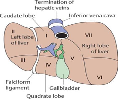
Caudate lobe (segment I) is a physiological independent part of liver, supplied by both right and left hepatic arteries; right and left branches of portal vein and drains bile into both right and left hepatic duct.
Weight of liver ‑
| A |
600-800 gm |
|
| B |
1000-1200gm |
|
| C |
1400-1600gm |
|
| D |
1800-2000gm |
Weight of liver ‑
| A |
600-800 gm |
|
| B |
1000-1200gm |
|
| C |
1400-1600gm |
|
| D |
1800-2000gm |
Ans. is ‘c’ i.e., 1400-1600 gm
- Liver is the largest gland of body situated in right upper quadrant of abdominal cavity and occupies whole of the right hypochondrium, greater part of epigastrium with extension into left hypochondrium.
- It weighs about 1500-1600 gm in males and 1200-1300 gm in females.
- Liver has five surfaces : anterior, posterior, superior, inferior and right.
- It has only one prominent border, inferior border.
- Liver is covered by Glisson’s capsule.
- Interior of liver is divided into hexagonal lobules.
- Lobule contains sinusoids which have fenesterated endothelium covering the subendothelial space of Disse.
- Two important cells in liver are hepatocytes (Parenchymal cells) and kupffer cells (monocytic-mocrophages).
Caudate lobe of liver is ‑
| A |
I |
|
| B |
III |
|
| C |
IV |
|
| D |
VI |
Caudate lobe of liver is ‑
| A |
I |
|
| B |
III |
|
| C |
IV |
|
| D |
VI |
Ans. is ‘a’ i.e., I
Type of collagen found in space of Disse in liver is –
| A |
Collagen I & II |
|
| B |
Collagen III & IV |
|
| C |
Collagen II & |
|
| D |
Collagen II & V |
Type of collagen found in space of Disse in liver is –
| A |
Collagen I & II |
|
| B |
Collagen III & IV |
|
| C |
Collagen II & |
|
| D |
Collagen II & V |
Ans. is ‘b’ i.e., Collagen III & IV
