

MUSCLES OF MASTICATION
All of the following muscles are grouped together as “muscles of mastication”, EXCEPT?
| A |
Buccinator |
|
| B |
Masseter |
|
| C |
Temporalis |
|
| D |
Pterygoids |
All of the following muscles are grouped together as “muscles of mastication”, EXCEPT?
| A |
Buccinator |
|
| B |
Masseter |
|
| C |
Temporalis |
|
| D |
Pterygoids |
The muscles of mastication are masseter, temporalis, medial and lateral pterygoid. These muscles are innervated by the mandibular division of trigeminal nerve.
- Masseter is involved in elevation and protrusion of the mandible.
- Temporalis elevates the mandible, chiefly with its vertical fibers, retracts the protruded mandible with its horizontal posterior fibers. Its unilateral contraction helps in mastication.
- Medial pterygoid elevates the mandible.
- Lateral pterygoid: Its bilateral contraction initiates opening of the mouth by protruding the mandible and moving the articular disk forward. Its unilateral contraction elevates the mandible to the opposite side during mastication.
Branches from the Anterior division of the Mandibular nerve supplies all of the following muscles, EXCEPT?
| A |
Lateral Pterygoid |
|
| B |
Medial Pterygoid |
|
| C |
Masseter |
|
| D |
Temporalis |
Branches from the Anterior division of the Mandibular nerve supplies all of the following muscles, EXCEPT?
| A |
Lateral Pterygoid |
|
| B |
Medial Pterygoid |
|
| C |
Masseter |
|
| D |
Temporalis |
Branches from the anterior division of the mandibular nerve are the buccal nerve, the nerve to masseter, the deep temporal nerves to the temporalis muscle and the nerve to the lateral pterygoid. The nerve to the medial pterygoid is a branch from the main trunk of the mandibular nerve.
Which of the following muscles is not supplied by mandibular nerve?
| A |
Masseter |
|
| B |
Buccinator |
|
| C |
Tensor veli palati |
|
| D |
Posterior belly of digastric |
Which of the following muscles is not supplied by mandibular nerve?
| A |
Masseter |
|
| B |
Buccinator |
|
| C |
Tensor veli palati |
|
| D |
Posterior belly of digastric |
Mandibular nerve supplies muscles of mastication, mylohyoid, anterior belly of digastric, tensor tympani and tensor veli palati. Nerve to the medial pterygoid is a small branch of mandibular nerve that supplies the medial pterygoid muscle. It gives off two branches which pass without interruption through the otic ganglion to supply tensor tympani and tensor veli palati.
The muscles acting upon the temporomandibular joint for various actions are given below, EXCEPT?
| A |
Temporalis muscle |
|
| B |
Occipitofrontalis |
|
| C |
Masseter muscle |
|
| D |
Medial pterygoid muscle |
The muscles acting upon the temporomandibular joint for various actions are given below, EXCEPT?
| A |
Temporalis muscle |
|
| B |
Occipitofrontalis |
|
| C |
Masseter muscle |
|
| D |
Medial pterygoid muscle |
- Temporalis muscle: The temporalis muscle elevates the mandible.
- Masseter muscle: It elevates the mandible. It works functionally with the temporalis and pterygoid muscles to move the mandible at the TMJ, it is a muscle of mastication.
- Lateral pterygoid muscle: Contraction causes the mandibular condyle and the articular disc to move anteriorly, resulting in both protraction and depression of the mandible. It works synergistically with the medial pterygoid muscle to move the mandible from side to side.
- Medial pterygoid muscle: It elevates the mandible and moves it from side to side.
Which is the nucleus of Masseteric Reflex?
| A |
Superior sensory nucleus of trigeminal nerve |
|
| B |
Spinal nucleus of trigeminal nerve |
|
| C |
Mesencephalic nucleus of trigeminal nerve |
|
| D |
Dorsal nucleus of vagus nerve |
Which is the nucleus of Masseteric Reflex?
| A |
Superior sensory nucleus of trigeminal nerve |
|
| B |
Spinal nucleus of trigeminal nerve |
|
| C |
Mesencephalic nucleus of trigeminal nerve |
|
| D |
Dorsal nucleus of vagus nerve |
The reflex arc of masseteric reflex involves two nuclei of the trigeminal nerve, namely, the mesencephalic nucleus and the motor nucleus of trigeminal nerve .
Muscle which helps in opening of the mouth:
| A |
Lateral pterygoid |
|
| B |
Medial pterygoid |
|
| C |
Temporalis |
|
| D |
Masseter |
Muscle which helps in opening of the mouth:
| A |
Lateral pterygoid |
|
| B |
Medial pterygoid |
|
| C |
Temporalis |
|
| D |
Masseter |
Lateral Pterygoid
- The primary function of the lateral pterygoid muscle is to pull the head of the condyle out of the mandibular fossa along the articular eminence to protrude the mandible.
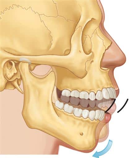
- A concerted effort of the lateral pterygoid muscles acts in helping lower the mandible and open the jaw whereas unilateral action of a lateral pterygoid produces contralateral excursion, usually performed in concert with the medial pterygoids
- Unlike the other three muscles of mastication, the lateral pterygoid is the only muscle of mastication that assists in depressing the mandible (opening the jaw).
- At the beginning of this action it is assisted by the digastric,mylohyoid and geniohvoid muscles.
Elevation of jaw is done by all except:
| A |
Temporalis |
|
| B |
Masseter |
|
| C |
Lateral pterygoids |
|
| D |
Medial pterygoids |
Elevation of jaw is done by all except:
| A |
Temporalis |
|
| B |
Masseter |
|
| C |
Lateral pterygoids |
|
| D |
Medial pterygoids |
Depression of the Mandible
Depression of the mandible is brought about by contraction of the digastrics, the geniohyoids, and the mylohyoids; the lateral pterygoids play an important role by pulling the mandible forward.
Elevation of the Mandible
Elevation of the mandible is brought about by contraction of the temporalis, the masseter, and the medial pterygoids. The head of the mandible is pulled backward by the posterior fibers of the temporalis.
Protrusion of the Mandible
In protrusion, the lower teeth are drawn forward over the upper teeth, which is brought about by contraction of the lateral pterygoid muscles of both sides, assisted by both medial pterygoids.
Refraction of the Mandible
The articular disc and the head of the mandible are pulled backward into the mandibular fossa. Retraction is brought about by contraction of the posterior fibers of the temporalis.
Lateral Chewing Movements
These are accomplished by alternately protruding and retracting the mandible on each side. For this to take place, a certain amount of rotation occurs, and the muscles responsible on both sides work alternately like turning the chin to left side produced by left lateral pterygoid and right medial pterygoid and vice versa.
Muscle which helps in this moment of the mandible
| A |
Lateral pterygoid |
|
| B |
Medial pterygoid |
|
| C |
Temporalis |
|
| D |
Masseter |
Muscle which helps in this moment of the mandible
| A |
Lateral pterygoid |
|
| B |
Medial pterygoid |
|
| C |
Temporalis |
|
| D |
Masseter |
Lateral Pterygoid
- The primary function of the lateral pterygoid muscle is to pull the head of the condyle out of the mandibular fossa along the articular eminence to protrude the mandible.
The muscles acting upon the structure encircled in picture for various actions are given below, EXCEPT

| A |
Temporalis muscle |
|
| B |
Occipitofrontalis |
|
| C |
Masseter muscle |
|
| D |
Medial pterygoid muscle |
The muscles acting upon the structure encircled in picture for various actions are given below, EXCEPT
| A |
Temporalis muscle |
|
| B |
Occipitofrontalis |
|
| C |
Masseter muscle |
|
| D |
Medial pterygoid muscle |
- Temporalis muscle: The temporalis muscle elevates the mandible.
- Masseter muscle: It elevates the mandible. It works functionally with the temporalis and pterygoid muscles to move the mandible at the TMJ, it is a muscle of mastication.
- Lateral pterygoid muscle: Contraction causes the mandibular condyle and the articular disc to move anteriorly, resulting in both protraction and depression of the mandible.
- Medial pterygoid muscle: It elevates the mandible and moves it from side to side

