
Non union and Mal-union
The following fractures are known for nonunion, EXCEPT:
| A |
Fracture lower half of tibia |
|
| B |
Fracture of neck of femur |
|
| C |
Fracture of scaphoid |
|
| D |
Supracondylar fracture of humerus |
The following fractures are known for nonunion, EXCEPT:
| A |
Fracture lower half of tibia |
|
| B |
Fracture of neck of femur |
|
| C |
Fracture of scaphoid |
|
| D |
Supracondylar fracture of humerus |
The distal humeral metaphysis (supracondylar area) is a well vascularized area with remarkably rapid healing and nonunion of a supracondylar fracture is extremely rare. Non union is almost never seen without infection (open wounds) and/ or extensive soft tissue devascularization.
Pappu, 7 yrs old young boy, had fracture of lateral condyle of femur. He developed malunion as the fracture was not reduced anatomically. Malunion will produce:
| A |
Genu valgum |
|
| B |
Genu varum |
|
| C |
Genu recurvatum |
|
| D |
Dislocation of knee |
Pappu, 7 yrs old young boy, had fracture of lateral condyle of femur. He developed malunion as the fracture was not reduced anatomically. Malunion will produce:
| A |
Genu valgum |
|
| B |
Genu varum |
|
| C |
Genu recurvatum |
|
| D |
Dislocation of knee |
Due to decrease growth over the lateral aspect in relation to the medial aspect, the person may develop Genu valgum.
Ref: Essential Orthopedics By J Maheswari, 2nd Edition, Page 274
All of the following factors fascilitate non union except:
| A |
Haematoma formation |
|
| B |
Periosteal injuries |
|
| C |
Absence of nerve supply |
|
| D |
Chronic infection |
All of the following factors fascilitate non union except:
| A |
Haematoma formation |
|
| B |
Periosteal injuries |
|
| C |
Absence of nerve supply |
|
| D |
Chronic infection |
A i.e. Hematoma formation
Fracture healing process passes through several stages. In which the, first is stage of hematoma formationQ and the last is stage of remodelingQ
Fracture Healing
Bone is a unique tissue because it heals by the formation of normal bone, as opposed to scar tissue. Infact it is considered a nonunion when a bone heals by fibroblastic response. The majority of fractures heal by secondary (callus) healing through a combination of intramembranous – ossification (hard callus formation) & endochondral ossificaion (soft callus formation). Bone healing can be divided into following phases depending on the biological events.
* There are two sources of repair tissues (callus), medullary & periosteal. The former is most marked in cancellous hone, while the later predominates in diaphyseal fracture. Periosteal callus may be of two types – primary callus (from DOPC) & external bridging callus (from IOPC), its appearance is inhibited by rigid fixation as plate and is predominant form of healing in cast immobilization &
intramedullary nailing. (i.e. micromovements present)
• Late medullary callus is more dependent on intramedullary vascularity (not on external soft trissue) and is important in fractures immobilized by rigid plating (i.e. no micrmovements)
* Phenomenon of Creeping Substitution occurs in cancellous bone (i.e. blood vessels invading the trabeculae of bone directly causing bone apposition)
* The modification of previously undifferentiated soft tissue cells in to osteogenic cells (IOPC) is termed bone induction.

The most common cause of non union is
| A |
Infection |
|
| B |
Inadequate immobilization |
|
| C |
Ischaemia |
|
| D |
Soft tissue interposition |
The most common cause of non union is
| A |
Infection |
|
| B |
Inadequate immobilization |
|
| C |
Ischaemia |
|
| D |
Soft tissue interposition |
B i.e. Inadequate immobilization
Non – union is a complication of :
| A |
Scaphoid # |
|
| B |
Colle’s # |
|
| C |
Inter- trochanteric # of hip |
|
| D |
Supra condylor # of the humerus |
Non – union is a complication of :
| A |
Scaphoid # |
|
| B |
Colle’s # |
|
| C |
Inter- trochanteric # of hip |
|
| D |
Supra condylor # of the humerus |
A i.e. Scaphoid Fracture
Fracture head & neck femur, fracture body of talus and fractures through waist of scaphoid most commonly leads to nonunion and avascular necrosis due to poor blood supplyQ
The most common complication of clavicle fracture is
| A |
Injury to brachial plexus |
|
| B |
Malunuion |
|
| C |
Stiffness of shoulder |
|
| D |
Non union |
The most common complication of clavicle fracture is
| A |
Injury to brachial plexus |
|
| B |
Malunuion |
|
| C |
Stiffness of shoulder |
|
| D |
Non union |
B i.e. Malunion
The most important cause of Nonunion of fracture of – humeral shaft is
| A |
Comminuted fracture |
|
| B |
Compound (Open) fracture |
|
| C |
Overriding of fracture ends |
|
| D |
Distraction at fracture site |
The most important cause of Nonunion of fracture of – humeral shaft is
| A |
Comminuted fracture |
|
| B |
Compound (Open) fracture |
|
| C |
Overriding of fracture ends |
|
| D |
Distraction at fracture site |
D i.e. Distraction at fracture site
Fracture shaft humerus is the easiest of major long bone fractures to be treated by conservative methods. And the most common cause of delayed union or nonunion is distraction at fracture siteQ due to gravity and weight of plaster.
Which of the following is known for Non union in children, if left untreated?
| A |
lntercondylar fracture of humerus |
|
| B |
Fracture shaft of humerus |
|
| C |
Fracture shaft of femur |
|
| D |
Fracture distal 1/3rd of tibia |
Which of the following is known for Non union in children, if left untreated?
| A |
lntercondylar fracture of humerus |
|
| B |
Fracture shaft of humerus |
|
| C |
Fracture shaft of femur |
|
| D |
Fracture distal 1/3rd of tibia |
B i.e. Fracture of shaft humerus
Growth disturbance, nonunion, elbow instability & late ulnar nerve palsy is commonly seen in
| A |
Fracture supracondylar humerus |
|
| B |
Fracture medial condyle |
|
| C |
Fracture lateral condyle |
|
| D |
Fracture head radius |
Growth disturbance, nonunion, elbow instability & late ulnar nerve palsy is commonly seen in
| A |
Fracture supracondylar humerus |
|
| B |
Fracture medial condyle |
|
| C |
Fracture lateral condyle |
|
| D |
Fracture head radius |
C i.e. Fracture lateral condyle
Milch Classification of Fracture Lateral Condyle Humerus
Mitch type I (Salter Harris type IV)
– Less common type
– Fracture running through the secondary ossification centre of capitullum and entering the joint lateral to capitulotrochlear groove Cause growth defectQ
Mitch type II (Salter Harris type II (?) IV (?))
– Commonest
– Fracture starting in metaphysis and running along the physis of lateral condyle into trochlear i.e. fracture extends medial to capitulotrochlear groove.
– Make ulnar
– humeral (elbow) joint unstableQ.
* If the lateral condyle is left capsized nonunion is inevitableQ : with growth elbow becomes increasingly valgus and tardy ulnar nerve palsyQ is then likely to develop.
A 10-year-old boy presenting with a cubitus varus deformity and a history of trauma 3 months back on clinical examination, has the preserved 3 bony point relationship of the elbow. The most probable diagnosis is
| A |
Old unreduced dislocation of elbow |
|
| B |
Non-union lateral condylar fracture of humerus |
|
| C |
Malunited intercondylar fracture of humerus |
|
| D |
Malunited supracondylar fracture of humerus |
D i.e. Malunited Supracondylar fracture
The malunion of supracondylar fracture of the humerus most commonly leads to:
| A |
Flexion deformity |
|
| B |
Cubitus varus |
|
| C |
Cubitus valgus |
|
| D |
Extension deformity |
The malunion of supracondylar fracture of the humerus most commonly leads to:
| A |
Flexion deformity |
|
| B |
Cubitus varus |
|
| C |
Cubitus valgus |
|
| D |
Extension deformity |
B i.e. Cabitus Varus
The most common complication of supracondylar fracture is
| A |
Osteosarcoma |
|
| B |
Genu valgum |
|
| C |
Blood vessel injury |
|
| D |
Malunion with gun stock deformity |
The most common complication of supracondylar fracture is
| A |
Osteosarcoma |
|
| B |
Genu valgum |
|
| C |
Blood vessel injury |
|
| D |
Malunion with gun stock deformity |
D i.e. Malunion with gunstock deformity
The following fractures are known for Non-union except:
| A |
Fracture of lower half of tibia |
|
| B |
Fracture of neck of femur |
|
| C |
Fracture of scaphoid |
|
| D |
Supracondylar fracture of humerus |
The following fractures are known for Non-union except:
| A |
Fracture of lower half of tibia |
|
| B |
Fracture of neck of femur |
|
| C |
Fracture of scaphoid |
|
| D |
Supracondylar fracture of humerus |
D i.e. Supracondylar humerus
The distal humeral metaphysis (supra condylar area) is a well vascularized area with remarkably rapid healing and nonunion of a supracondylar fracture is extremely rareQ. Nonunion is almost never seen without infection (open wounds) and /or extensive soft tissue devascularization.
A 10-year-old boy presenting with a cubitus varus deformity and a history of trauma 3 months back on clinical examination, has the preserved 3 bony point relationship of the elbow. The most probable diagnosis is
| A |
Old unreduced dislocation of elbow |
|
| B |
Non-union lateral condylar fracture of humerus |
|
| C |
Malunited intercondylar fracture of humerus |
|
| D |
Malunited supracondylar fracture of humerus |
D i.e. Malunited supracondylar humerus
Cubitus varus deformity and preservation of three point bony relationQ favours the diagnosis of malunited fracture supracondylar humrus.
Commonest complication of Colles’ fracture is:
| A |
Nonunion |
|
| B |
Malunion |
|
| C |
Vascular injury |
|
| D |
Sudeck’s osteodystrophy |
Commonest complication of Colles’ fracture is:
| A |
Nonunion |
|
| B |
Malunion |
|
| C |
Vascular injury |
|
| D |
Sudeck’s osteodystrophy |
B i.e. Malunion
In nonunion of scaphoid vescularized muscle pedicle graft is taken from.
| A |
Pronator teris |
|
| B |
Brachioradialis |
|
| C |
Pronator quadratus |
|
| D |
Extensor pollicis longus |
In nonunion of scaphoid vescularized muscle pedicle graft is taken from.
| A |
Pronator teris |
|
| B |
Brachioradialis |
|
| C |
Pronator quadratus |
|
| D |
Extensor pollicis longus |
C i.e. Pronator quadrates
Blood supply of scaphoid enters distally mainly through the dorsal ridge (spiral groove, narrow non articular region in the waist) and proximal segment is supplied by retrograde intraosseous blood flow from distal to proximal. This unusual pattern of vascularity is responsible for higher probability of nonunion & avascular necrosis of proximal fragment in fractures of scaphoid. Therefore 0-1% of distal third, 20% of middle third, 40% of proximal third and 100% of proximal pole fractures result in AVN and nonunion.
Scaphoid Fracture
Functional Anatomy & Epidemiology
Scaphoid is the most commonly fractured bone in the – carpusQ, in adult as well as children. However unlike in adults and adolescents, the fracture is rare in young children. (d/t cartilagenous nature of carpal bones in children)
Scaphoid fracture is seen most commonly in males between the ages of 15 and 30. (adolscents & adults)Q
– Scaphoid may be divided into proximal, middle and distal thirds. The middle third is termed the waist. The scaphoid tubercle forms the distal volar prominence. Middle third (Waist) fractures are most commonQ accounting for – 70% of scaphoid fractures > proximal pole fracture (20%) > distal pole fracture (10%), in adults & adolescents.
Investigation
Distal pole avulsion type fracture is most common fracture type in children Q.
Anteroposterior, lateral and oblique views are all essential; often a recent fracture shown only in the oblique viewQ. So a routine scaphoid x- ray series must include:-
Radial and ulnar deviaton PA views with the wrist in about 30° of extension (by asking to make a fist gently)
- True lateral view
- Radial oblique (supinated PA) view and ulnar oblique (pronated PA) view.
- Comparison view of opposite wrist.
However it is common for the fracture not to be visible on the initial films. If doubt exists, the scaphoid should be immobilized in plaster and repeat radiograph obtained in 1421 days as the late films are usually positiveQ.
Bone scan, trispiral tomography are other investigations ; occult scaphoid fractures can be reliably diagnosed by MRI & nucleotide scan.
Vascularity, Nonunion & AVN
Blood supply of scaphoid is precarious. It receives most of its blood supply from two major vascular pedicles (dorsal & volar branches) from the radial artery, both entering distallyQ.
Volar (palmar) scaphoid branch of radial artery enters the scaphoid tubercle and supplies its distal 20-30%.
Dorsal scaphoid branch of radial artery enters through numerous small foramina along the spiral groove & dorsal ridge and account for 80% of its blood supply.
There is usually no or rarely a single perforater proximal to the waist of scaphoid. Hence vascularity of proximal fragment is maintained by retrograde intraosseous blood flow from distal to proximal; 7080% of which is provided by dorsal branch. The distal scaphoid has a dense vascular network whereas intraosseous vascular density declines proximally, leaving the proximal pole with sparse blood supply. Because of this unusual retrograde vascular supply to proximal segment, the scaphoid has a high risk of nonunion and AVN after fracture through waist and proximal pole.
Because the scaphoid articulates with four carpal bones & radius, most of its surface is composed of articular
cartilage. Therefore, the vascular supply comes through -a narrow non articular region in the waist. Most of the blood supply to the scaphoid enters distally, so blood supply of scaphoid diminishes proximallyQ. This accounts for the fact that 1% of distal third, 20% of middle third, 40% of proximal third and 100% of proximal pole fractures result in avascular necrosis or non union of the proximal fragment2.
–Because it articulates with distal radius, and with 4 of remaining 7 carpal bones, the scaphoid moves with nearly all carpal motions, especially volar flexion. Any alteration of its articular surface through fracture, dislocation, or subluxation or any change of its stability by ligamentous rupture can cause severe secondary changes throughout the entire carpus.
Management
Stable and undisplaced fractures are treated by scaphoid cast immobilization (glass holding dorsal and radial flexion positionQ impacts the scaphoid fragments and minimises the shearing effects), for 4 – 8 weeks in distal third, 6 – 12 weeks in middle third and 12- 20 weeks in proximal third fractures. Scaphoid cast is applied from the upper forearm to just short of meta carpophalyngeal joints of fingers, but incorporating proximal phalynx of thumb. The wrist is held dorsiflexed and the thumb forwards in “glass holding position”
– A displaced fracture, by definition, is one with > 1 mm of step-off or >60°of scapholunate or >15° of lunato- capitate angulation. The subtle signs of displacement or instability are – opening & obliquity of fracture line, angulation of distal fragment, and fore shortening of scaphoid image. Displaced- unstable fractures are treated by percutaneous screw fixation or OR & IF by k – wires, compression screw or Herbert screw.
– Undisplaced ununited (nonunion) fractures may be treated by excavation of the scaphoid & placement of volar inlay corticocancellous bone graft (Matti Russe procedure). In most cases of stable nonunion cancellous bone graft from either the distal radius (for small defects) or the iliac crest (preferable because of its superior osteogenic and mechanical properties) is packed into the defect. Dorsal cartilage is not disturbed. This provides a hinge and facilitates assessment of scaphoid length. If fracture site is angulated or collapsed cortico cancellous volar graft is employed to correct the deformity.
If the proximal pole is avascular and no significant radiocarpal arthritis is present, revascularization of the scaphoid with a vascularized bone graft from dorsal radius or preferably pronator quadratus graftQ should be performed.
Partial radial stylodectomy should be performed in all patients with radiological signs of stage I radioscaphoid arthritis limited to scaphoid and radial styloid.
– Once degenerative arthritis is evident at the radio -carpal joint, salvage procedures includes proximal row carpectomy, scaphoid excision and mid carpal arthodesis or total wrist arthrodesis.
Clinical Presentation
– The patient is usually and adolescent boy or young adult who gives a h/o falling on outstretched hand usually during active sports. Commonly the injury is misinterpreted as “just a sprain”. Like colle’s it is a supination – dorsiflexion injury.
– Fullness & tenderness in anatomical snuff boxQ. Radial side wrist pain; passive dorsiflexion to the radial side is painful, grip is weak and release of grip gives transitory pain, resisted pinch between the thumb and index finger is uncomfortable. Proximal pressure along the axis of the thumb may be painfulQ.
Nonunion is a very common complication of intracapsular fractures of the neck of femur. Which of the following is not a very important cause for the same?
| A |
Inadequate immobilization |
|
| B |
Inadequate blood supply |
|
| C |
Inhibitory effect of synovial fluid |
|
| D |
Stress at fracture site due to muscle spasm |
D i.e. Stress at fracture site due to muscle spasm
Commonest complication of extra capsular intertrochanteric fracture of neck of femur is:
| A |
Non union |
|
| B |
Ischemic necrosis |
|
| C |
Malunion |
|
| D |
Pulmonary complications |
Commonest complication of extra capsular intertrochanteric fracture of neck of femur is:
| A |
Non union |
|
| B |
Ischemic necrosis |
|
| C |
Malunion |
|
| D |
Pulmonary complications |
C i.e. Malunion
The most common complication of intracapsular fracture neck of femur is
| A |
Mal union |
|
| B |
Osteoarthritis |
|
| C |
Non-Union |
|
| D |
Shortening |
The most common complication of intracapsular fracture neck of femur is
| A |
Mal union |
|
| B |
Osteoarthritis |
|
| C |
Non-Union |
|
| D |
Shortening |
C i.e. Nonunion
Treatment of Non-union of # shaft femur
| A |
Open reduction with external fixation |
|
| B |
Excision of the bone |
|
| C |
Bone grafting with internal fixation with K-Nail |
|
| D |
All of the following |
Treatment of Non-union of # shaft femur
| A |
Open reduction with external fixation |
|
| B |
Excision of the bone |
|
| C |
Bone grafting with internal fixation with K-Nail |
|
| D |
All of the following |
C i.e. Bone grafting with internal fixation with K-Nail
Best treatment of 3 weeks old, fracture shaft femur with nonunion is
| A |
Bone graft with internal fixation |
|
| B |
External fixation |
|
| C |
Internal fixation only |
|
| D |
Prosthesis |
Best treatment of 3 weeks old, fracture shaft femur with nonunion is
| A |
Bone graft with internal fixation |
|
| B |
External fixation |
|
| C |
Internal fixation only |
|
| D |
Prosthesis |
A i.e. Bone grafting with internal fixation
Non union is a common feature of fracture of
| A |
Supracondylar humerus |
|
| B |
Clavicle |
|
| C |
Lower tibia |
|
| D |
Coracoid process |
Non union is a common feature of fracture of
| A |
Supracondylar humerus |
|
| B |
Clavicle |
|
| C |
Lower tibia |
|
| D |
Coracoid process |
C i.e. Lower tibia
Fracture through the lower third of tibia. is more liable to go onto delayed union because the lower fragment becomes relatively avascular dlt poor vascularityQ.
Commonest complication of extra capsular fracture of femur is:
September 2005
| A |
Non union |
|
| B |
Mal union |
|
| C |
Avascular necrosis |
|
| D |
Osteoarthritis |
Commonest complication of extra capsular fracture of femur is:
September 2005
| A |
Non union |
|
| B |
Mal union |
|
| C |
Avascular necrosis |
|
| D |
Osteoarthritis |
Ans. B: Mal Union
Complications of extra-capsular/ inter-trochanteric fracture:
- Union of the fracture in an unacceptable position resulting in a deformity/ malunion- the fracture heals with unacceptable shortening, rotation, and/or angulation of the extremity, resulting in decreased mobility and subsequent handicap, impairment, and disability.
- Rarely osteo-arthritis.
Common sites of fracture non union are the following except –
| A |
Waist of scaphoid |
|
| B |
Neck of femur |
|
| C |
Distal 1/3 tibia fibula |
|
| D |
Distal end radius |
Common sites of fracture non union are the following except –
| A |
Waist of scaphoid |
|
| B |
Neck of femur |
|
| C |
Distal 1/3 tibia fibula |
|
| D |
Distal end radius |
Ans. is `d’ i.e., Distal end radius
Non-union
- When a fracture fails to unit completely even after the stipulated time, it is termed as non-union.
- Both, In non-union and delayed union, fracture does not unite in the usual stipulated time. The difference between two is that
- In delayed union, union occurs but at a slower rate, i.e. there is presence of clinical and radiological progressive signs of union though at a slower rate. Healing may occur without surgical intervention by just prolonging the duration of immobilization.
- In non-union, union does not occur at all, i.e. there is complete cessation of healing process in which fibrous tissue is never replaced by bony matrix. So, there is absence of clinical and radiological progressive signs of union. Fracture healing will not occur without surgical intervention, e.g. bone grafting, illizarov’s technique.
- Common sites of non-union are neck of femur, scaphoid, lower third of tibia, lateral condyle of humerus and lower third of ulna.
|
True statement regarding Fracture of the bone shown in this image |

| A |
Most common Complication is Malunion |
|
| B |
Occurs at the jn.of medial 1/3rd & lateral 2/3r |
|
| C |
Usually occurs due to fall on elbow |
|
| D |
Communitted fracture is common |
|
True statement regarding Fracture of the bone shown in this image |
| A |
Most common Complication is Malunion |
|
| B |
Occurs at the jn.of medial 1/3rd & lateral 2/3r |
|
| C |
Usually occurs due to fall on elbow |
|
| D |
Communitted fracture is common |
A i.e. Most common complication is malunion.
Clavicle Bone is shown in the image.
Peculiar features of clavicle
- First bone to ossify in the body.
- Only long bone which ossify from two primary and one secondary centre.
- Only long bone in the body lying horizontal.
- Only long bone which ossify from a membrane (Intramembranous ossification).
- Only link between the appendicular and the axial skeleton.
- The most common bone to be fractured in children and during birth.
- Only long bone subcutaneous throughout.
- It has no medullary cavity.
- The most common site for fracture of clavicle is the junction of the middle third and outer third (i.e. junction of medial 2/3rd and lateral 1/3rd).
- Most common complication of Clavicular fracture is Malunion
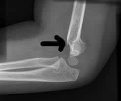
A 10-year-old boy presenting with a condition shown in the picture below had a history of trauma 3 months back. On clinical examination, has the preserved 3 bony point relationship of the elbow. The most probable diagnosis is ?

| A |
Old unreduced dislocation of elbow. |
|
| B |
Non-union lateral condylar fracture of humerus. |
|
| C |
Malunited intercondylar fracture of humerus. |
|
| D |
Malunited supracondylar fracture of humerus. |
A 10-year-old boy presenting with a condition shown in the picture below had a history of trauma 3 months back. On clinical examination, has the preserved 3 bony point relationship of the elbow. The most probable diagnosis is ?
| A |
Old unreduced dislocation of elbow. |
|
| B |
Non-union lateral condylar fracture of humerus. |
|
| C |
Malunited intercondylar fracture of humerus. |
|
| D |
Malunited supracondylar fracture of humerus. |
Cubitus varus deformity as shown in the picture above and preservation of three point bony relation favours the diagnosis of malunited fracture supracondylar humrus.
Complications of the fracture shown in the picture below are all except ?
| A |
Elbow stiffness. |
|
| B |
Mal union. |
|
| C |
Non union. |
|
| D |
Myositis ossification. |
Complications of the fracture shown in the picture below are all except ?
| A |
Elbow stiffness. |
|
| B |
Mal union. |
|
| C |
Non union. |
|
| D |
Myositis ossification. |
The fracture shown in the picture above represents supracondylar fracture of humerus.
Complication is non union.
Complication of neck femur fracture are all except‑
| A |
Nonunion |
|
| B |
Malunion |
|
| C |
AVN |
|
| D |
Osteoarthritis |
Complication of neck femur fracture are all except‑
| A |
Nonunion |
|
| B |
Malunion |
|
| C |
AVN |
|
| D |
Osteoarthritis |
Ans. is ‘b’ i.e., Malunion
Complications of femoral neck fracture
- Fractures of the neck of the femur are more prone to serious complications than in any other fracture. All the complications affect fractures with displacement rather than impacted abducted (valgus impacted) fractures. The important complications are :
1. Avascular necrosis of femoral head
- AVN is the most common complication of femoral neck fracture.
- It occurs in 15-35% of cases of displaced fractures and
2. Non-union
- Non-union is the second most common complication of femoral neck fracture.
- It occurs in 10-30% of cases of displaced fractures and
3. Secondary osteoarthritis
- It occurs a few years following fracture neck femur.
- Avascular necrosis or collapse of femoral head leads to secondary osteoarthritis of the hip joint.
Mc Murray’s osteotomy is done for-
| A |
Malunited intertrochantric fracture of femur |
|
| B |
Nonunion transcervical neck fracture of femur |
|
| C |
Nonunion lateral condyle fracture of humerus |
|
| D |
Malunited supracondylar fracture of humerus |
Mc Murray’s osteotomy is done for-
| A |
Malunited intertrochantric fracture of femur |
|
| B |
Nonunion transcervical neck fracture of femur |
|
| C |
Nonunion lateral condyle fracture of humerus |
|
| D |
Malunited supracondylar fracture of humerus |
Ans. is ‘b’ i.e., Non-union transcervical neck fracture of femur
Fracture neck femur cause of non-union‑
| A |
Injury to blood supply with shearing stress |
|
| B |
Poor nutrition of the patient |
|
| C |
Smoking |
|
| D |
Old age and osteoporosis |
Fracture neck femur cause of non-union‑
| A |
Injury to blood supply with shearing stress |
|
| B |
Poor nutrition of the patient |
|
| C |
Smoking |
|
| D |
Old age and osteoporosis |
Ans. is ‘a’ i.e., Injury to blood supply with shearing stress
- Causes of non-union in fracture neck of femur are :‑
- Fracture morphology – high fracture angle and increased shear.
- Displaced fracture grade 3/4.
- Fracture comminution.
- Inadequate reduction and stability of fixation.
- Poor bone quality.
- Injury to vascularity- direct and tamponade effect.
- Absence of cambium layer in periosteum.
- Factors in synovial fluid which inhibit the callus formation.
- Lack of hematoma.
- Washing away and dilution of osteogenic factors.
