

OBTURATOR NERVE
Which muscles are supplied by obturator nerve‑
| A |
Abductor muscle |
|
| B |
Adductor compartment |
|
| C |
Extensor compartment |
|
| D |
Flexor compartment |
Which muscles are supplied by obturator nerve‑
| A |
Abductor muscle |
|
| B |
Adductor compartment |
|
| C |
Extensor compartment |
|
| D |
Flexor compartment |
B. i.e. Adductor compartment
Adductor compartment of thigh is supplied by obturator nerveQ; and anterior flexor compartmentof thigh by femoral nerve. Gluteus medius, minimus & tensor fascia lata are supplied by superior gluteal nerve, (Mn- “Middle & Minimum class Tension is superior”). Where as gluteus maximus is supplied by inferior gluteal nerve.
Nerve supply of adductor magnus is through:
| A |
Tibial part of sciatic nerve |
|
| B |
Obturator nerve |
|
| C |
Both |
|
| D |
None |
Nerve supply of adductor magnus is through:
| A |
Tibial part of sciatic nerve |
|
| B |
Obturator nerve |
|
| C |
Both |
|
| D |
None |
Innervation of adductor magnus
- Posterior division of obturator nerve innervates most of the adductor magnus
- Vertical or hamstring portion innervated by tibial nerve (L2, L3, L4)
Nerve Supply of the Muscle marked as Muscle A in the diagram is

| A |
Tibial part of sciatic nerve |
|
| B |
Obturator nerve |
|
| C |
Both |
|
| D |
None |
Nerve Supply of the Muscle marked as Muscle A in the diagram is
| A |
Tibial part of sciatic nerve |
|
| B |
Obturator nerve |
|
| C |
Both |
|
| D |
None |
Innervation of adductor magnus
- Posterior division of obturator nerve innervates most of the adductor magnus
- Vertical or hamstring portion innervated by tibial nerve (L2, L3, L4)
All nerves pass thorugh greater sciatic notch except ‑
| A |
Superior gluteal nerve |
|
| B |
Inferior gluteal nerve |
|
| C |
Sciatic nerve |
|
| D |
Obturator nerve |
All nerves pass thorugh greater sciatic notch except ‑
| A |
Superior gluteal nerve |
|
| B |
Inferior gluteal nerve |
|
| C |
Sciatic nerve |
|
| D |
Obturator nerve |
Ans. is ‘d’ i.e., Obturator nerve
Structures crossing dorsal surface of the given structure marked by a “red arrow” are ? 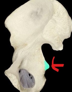
| A |
Internal pudendal vessel. |
|
| B |
Pudendal nerve. |
|
| C |
Obturator nerve. |
|
| D |
Nerve to obturator internus. |
Structures crossing dorsal surface of the given structure marked by a “red arrow” are ?
| A |
Internal pudendal vessel. |
|
| B |
Pudendal nerve. |
|
| C |
Obturator nerve. |
|
| D |
Nerve to obturator internus. |
Ans;C).Obturator nerve
The structure marked by a red arrow represents the ischial spine.
ISCHIAL SPINE–a thin pointed triangular eminence that projects from the dorsal border of the ischium and gives attachment to the gemellus superior on its external surface and to the coccygeus, levator ani, and pelvic fascia on its internal surface
- The structure crossing dorsal surface of ischial spine is the Obturator nerve.
- Psoas major, iliacus & pectineus muscles, femoral vessels and nerve, femoral branch of genitofemoral nerve, lateral cutaneous nerve of thigh and lymphatics pass below inguinal ligament.
- ‘PIN’ structures i.e. Pudendal nerve, Internal Pudendal vessels, Nerve to obturator internus come out of greater sciatic foramen, cross the dorsal surface of ischial spine & enter into lesser sciatic foramen.
- From the posterior border of the body of the Ischium there extends backward a thin and pointed triangular eminence, the ischial spine, more or less elongated in different subjects.
Surfaces
| external surface | gives attachment to the Gemellus superior |
| internal surface | gives attachment to the Coccygeus, Levator ani, and the pelvic fascia |
| pointed extremity | the sacrospinous ligament is attached. |
It can serve as a landmark in pudendal anesthesia.

