
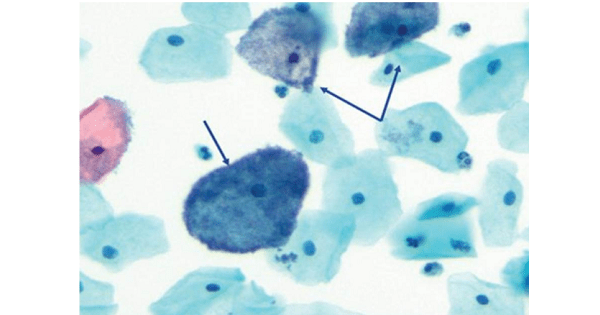
PAPANICOLAOU TEST
Pap smear is useful in the diagnosis of all EXCEPT :
| A |
Gonorrhoea |
|
| B |
Trichomonas vaginalis |
|
| C |
Human papilloma virus |
|
| D |
Inflammatory changes |
Pap smear is useful in the diagnosis of all EXCEPT :
| A |
Gonorrhoea |
|
| B |
Trichomonas vaginalis |
|
| C |
Human papilloma virus |
|
| D |
Inflammatory changes |
Gonorrhoea
Papsmear is a screening test for diagnosis of cervical Intraepithelial Neoplasia (CIN) and Carcinoma cervix.
Besides these it is also helpful in detecting :
- Trichomonas vaginalis° • Candidiasis°
- Bacterial vaginosis° • Actinomycosis°
- Herpes simplex virus infection° • Inflammatory changes
- Cellular changes associated with radiation • Cellular changes associated with intrauterine devices.
- Glandular cell status post hysterectomy • Atrophy (i.e., post surgery)
With respect to CIN and Ca cervix : Papsmear can diagnose epithelial cell abnormalities. (Both squamous and glandular).
|
Squamous cell abnormalities detected
|
Glandular cell abnormalities detected —1
|
It should be done in :
- All women > 20 years° (Harrison 17/e, p 491)
- All women 3 years after the onset of sexual activity°
- Women with unhealthy cervix°
- Women with intermenstrual bleeding or discharge°
- High risk cases like women exposed to early sexual life, multiple sexual partners, multipara or women with HIV, HPV or herpes simplex virus infection°
- Smokers.
All of the following are indications of doing PAP smear except :
| A |
> 20 years |
|
| B |
Pregnant female |
|
| C |
Sexually active female |
|
| D |
Menorrhagia |
All of the following are indications of doing PAP smear except :
| A |
> 20 years |
|
| B |
Pregnant female |
|
| C |
Sexually active female |
|
| D |
Menorrhagia |
Menorrhagia
Pap smear in pregnancy is :
| A |
Contraindicated |
|
| B |
Not useful |
|
| C |
Routine, as a part of screening |
|
| D |
Done in every patient |
Pap smear in pregnancy is :
| A |
Contraindicated |
|
| B |
Not useful |
|
| C |
Routine, as a part of screening |
|
| D |
Done in every patient |
Routine, as a part of screening
True about CIN
| A |
Premalignant lesion |
|
| B |
HPV predisposes |
|
| C |
Pap smear can detect it and Chlamydia infection can predispose both |
|
| D |
All |
True about CIN
| A |
Premalignant lesion |
|
| B |
HPV predisposes |
|
| C |
Pap smear can detect it and Chlamydia infection can predispose both |
|
| D |
All |
Ans. is a, b and c i.e. Premalignant lesion; HPV predisposes; Pap smear can detect it; Chlamydia infection can predispose; and Occurs at squamo-columnar junction
Cervical Intraepithelial Neoplasia (CIN) is the term used to describe the condition of cervix when a part or the full thickness of stratified squamous epithelial cells is replaced by dysplastic cells.
It is a premalignant condition.
CIN can be graded as
CIN I (Mild Dysplasia)
Dysplastic cells seen
in lower third of epithelial lining.
CIN II (Mod dysplasia) Dysplastic cells seen
in 2/3rd
of epithelial lining.
CIN III (Severe dysplasia
Dysplastic cells seen more than 2/3rd of epithelial lining.
Carcinoma in Situ
It represents full thickness dysmaturity, but basement membrane is intact.°
- Mild dysplasia can be seen in inflammatory conditions like Trichomoniasis and is reversible.
- CIN occurs at squamocolumnar Junction/ Transformation Zone.°
- Factors Predisposing to CIN / Ca cervix :
– Coitus before 18 years of age°
– Multiple sex partners°
– Delivery of lst baby before 20 years of age°
– Multiparity°
— Poor personal hygiene°
– Poor socioeconomic status°
– Smoking°, Alcohol, Drug abuse
– Women with : STD°, HIV°, HSV-2°, HPV infection° [16 (MC), 18, 31, 33, 35, 39, 45, 51, 52, 56, 58]
… Novak 14/e, p 568
– Immunosuppressed individuals°
– Women with H/0 Preinvasive lesion°
– Women on OCP°, Progesterone therapy for long time are predisposed to adenocarcinoma endocervix. From the above list it is clear that : STD’s predispose to CIN and chlamydia is an STD.
As far as specifically chlamydia is concerned.
“Infection with the herpes virus was previously thought to be the initiating event in cervical cancer; however,
infection with human papilloma virus (HPV) has now been determined to be the causal agent in the
development of cervical cancer, with herpes virus and Chlamydia trachomatis likely acting as cofactors.”
True about carcinoma in women :
| A |
Pap smear reduces incidences of Ca cervix |
|
| B |
Ca cervix is most common malignancyin Indian women |
|
| C |
Breast Ca is the most common malignacy in Indian women |
|
| D |
Next to Cervical Ca, lung carcinoma is next most common malignancy |
True about carcinoma in women :
| A |
Pap smear reduces incidences of Ca cervix |
|
| B |
Ca cervix is most common malignancyin Indian women |
|
| C |
Breast Ca is the most common malignacy in Indian women |
|
| D |
Next to Cervical Ca, lung carcinoma is next most common malignancy |
Ans. is a i.e. Pap smear reduces the incidences of Ca cervix
“The pap test has been successful in reducing the incidence of cervical cancer by 79% and mortality by 70% since 1950.” . So option ‘a ‘is correct.
Now option ‘b’says – Ca cervix is the most common malignancy in Indian women which is wrong because – Most common cancer in Indian females / Females worldwide = Breast cancer. …
Whereas most common genital cancer in Indian females = Ca cervix.
“Carcinoma of the cervix continues to be the most common genital cancer encountered in clinical practice in India. (80%).”
A 22-year-old woman comes to the physician for an annual examination. She has normal periods every month and has no complaints. She has no medical problems but does smoke one pack of cigarettes per day. She has intercourse with more than one partner. Examination is unremarkable, including a normal pelvic examination. A Papanicolaou smear shows a high- grade squamous intraepithelial lesion. Which of the following is the most appropriate next step in management?
| A |
Repeat Pap smear in 1 year |
|
| B |
Perform colposcopy |
|
| C |
Perform a cone biopsy |
|
| D |
Both A & B |
A 22-year-old woman comes to the physician for an annual examination. She has normal periods every month and has no complaints. She has no medical problems but does smoke one pack of cigarettes per day. She has intercourse with more than one partner. Examination is unremarkable, including a normal pelvic examination. A Papanicolaou smear shows a high- grade squamous intraepithelial lesion. Which of the following is the most appropriate next step in management?
| A |
Repeat Pap smear in 1 year |
|
| B |
Perform colposcopy |
|
| C |
Perform a cone biopsy |
|
| D |
Both A & B |
The Papanicolaou smear is an excellent screening technique for cervical cancer because it is easy to perform, has a relatively low-cost, and is noninvasive. All women who are either sexually active or older than 18 should have annual Pap smears. The downside of the Pap smear is that it has a low sensitivity and high false-negative rate. That is, many women with abnormal cervical cells will have a negative Pap smear. However, if a woman receives an annual Pap smear, it is likely that the lesion will be discovereD. Because the progression of cervical dysplasia to cancer takes time, it is believed that these lesions will be discovered early enough to cure them so long as annual screening occurs. This patient has what is called a high-grade squamous intraepithelial lesion (HGSIL). These lesions have a significant risk of eventually progressing to invasive cervical cancer if they are not treateD. Therefore, any patient with HGSIL on a Pap smear must undergo colposcopy with directed biopsies so that the lesion can be removeD. To repeat the Pap smear in 1 year ( Choice A)is incorrect. A 1- year follow-up is appropriate for a patient with normal Pap smears. This patient has HGSIL and therefore needs much closer follow-up. To repeat the Pap smear in 6 months (Choice B) is incorrect. A 6-month follow-up is appropriate for patients with a Pap smear showing atypical squamous cells of undetermined significance (ASCUS), or in some patients with a low-grade squamous intraepithelial lesion (LGSIL). To perform a cone biopsy (choice D. would be incorrect. Prior to surgical management of an abnormal Pap smear result, a tissue diagnosis should be made (Pap smear provides only a cytologic diagnosis).
Koilocytes with perinuclear halo on pap smear is pathognomic of?
| A | HPV infection | |
| B |
Metaplasia |
|
| C | Dysplasia | |
| D |
Bacterial vaginosis |
Koilocytes with perinuclear halo on pap smear is pathognomic of?
| A | HPV infection | |
| B |
Metaplasia |
|
| C | Dysplasia | |
| D |
Bacterial vaginosis |
HPV infection REF: Shaw’s Gynecology 13th ed p- 78, The pap test by Richard M. DeMay page 49-50
“Koilocytes, dyskeratocytes, macrocytes, etc. are some of the cytologic findings that are characteristic of condylomas (HPV). Koilocytes with perinuclear halo is pathognomonic of HPV infection”
|
Condition |
Type of cell |
|
Estrogen dominated uterus |
Normal cornified cells |
|
Progesterone dominated uterus |
Intermediate cells |
|
pregnancy |
Intermediate cells , Navicular cells |
|
menopausal |
Basal , parabasal |
|
HPV |
Koilocytes, dyskeratocytes, macrocytes with perinuclear halo. |
Koilocytes with dyskeratosis and perinuclear halo on pap smear is pathognomonic of which of the following?
| A |
HPV infection |
|
| B |
Metaplasia |
|
| C |
Dysplasia |
|
| D |
Bacterial vaginosis |
Koilocytes with dyskeratosis and perinuclear halo on pap smear is pathognomonic of which of the following?
| A |
HPV infection |
|
| B |
Metaplasia |
|
| C |
Dysplasia |
|
| D |
Bacterial vaginosis |
The presence of abnormal koilocytes (keratinocytes with pyknotic, deeply blue nuclei surrounded by a halo and clear cytoplasm with a paucity of keratohyaline granules), or koilocytosis, is a characteristic feature of productive HPV infection.
Other cytologic markers of HPV infection include acanthosis, dyskeratosis, and multinucleation.
Virus multiplication is confined to the host cell nucleus. Consequently, infected cells exhibit a high degree of nuclear atypia.
A Pap smear from a 30-year-old woman demonstrates protozoal parasites. These organisms are likely to be:
| A |
Cryptosporidium parvum |
|
| B |
Entamoeba histolytica |
|
| C |
Giardia lamblia |
|
| D |
Trichomonas vaginalis |
A Pap smear from a 30-year-old woman demonstrates protozoal parasites. These organisms are likely to be:
| A |
Cryptosporidium parvum |
|
| B |
Entamoeba histolytica |
|
| C |
Giardia lamblia |
|
| D |
Trichomonas vaginalis |
Trichomonas vaginalis (note “vagina” in the name) is the only organism listed to primarily affect the genital tract rather than the intestinal tract. The vaginitis it causes is characterized by a frothy, yellow discharge. The organism can be identified in Pap smears, or, more reliably, by special culture techniques that are becoming more widely available.
Infection in men is usually asymptomatic, but prostatitis and urethritis can also occur. Treatment with metronidazole is indicated for both the patient and the sexual partner(s). The other organisms listed cause gastrointestinal disease.
The physician takes a sample for Pap smear analysis from a 15 year old girl. Review of the Pap smear demonstrates the presence of koilocytes, which are approximately the same size as an intermediate cells, with enlarged hyperchromatic nuclei and perinuclear halos. The abnormal cells observed in the Pap smear specimen are infected by which of the following viruses?
| A |
AIDS virus |
|
| B |
Cytomegalovirus |
|
| C |
Herpes simplex l |
|
| D |
Human papillomavirus |
The physician takes a sample for Pap smear analysis from a 15 year old girl. Review of the Pap smear demonstrates the presence of koilocytes, which are approximately the same size as an intermediate cells, with enlarged hyperchromatic nuclei and perinuclear halos. The abnormal cells observed in the Pap smear specimen are infected by which of the following viruses?
| A |
AIDS virus |
|
| B |
Cytomegalovirus |
|
| C |
Herpes simplex l |
|
| D |
Human papillomavirus |
A 35 year old female presented with post coital bleeding. Next step of management is:
| A |
Pap smear and colposcopy |
|
| B |
Visual inspection with acetowhite |
|
| C |
Visual inspection with lugol’s iodine |
|
| D |
Colposcopy directed biopsy |
A 35 year old female presented with post coital bleeding. Next step of management is:
| A |
Pap smear and colposcopy |
|
| B |
Visual inspection with acetowhite |
|
| C |
Visual inspection with lugol’s iodine |
|
| D |
Colposcopy directed biopsy |
Postcoital bleeding (PCB) has many causes, including vaginitis, cervicitis, cervical dysplasia and malignancy, uterine lesions, and pregnancy.
Pap smear is an example of –
| A |
Primary level of prevention |
|
| B |
Secondary level of prevention |
|
| C |
Teritary level of prevention |
|
| D |
None of the above |
Pap smear is an example of –
| A |
Primary level of prevention |
|
| B |
Secondary level of prevention |
|
| C |
Teritary level of prevention |
|
| D |
None of the above |
Ans. is ‘b’ i.e., Secondary level of prevention
o In secondary prevention, action halts the progress of a disease at its incipient stage and prevents complication.
o Screening tests (e.g. – Pap smear), helps in diagnosis at early stage so that adequate treatment can halt disease progression and prevent complication.
Screening of cervical cancer at PHC level is done by?
| A |
History and clinical examination |
|
| B |
Colposcopy |
|
| C |
CT scan |
|
| D |
PAP smear |
Screening of cervical cancer at PHC level is done by?
| A |
History and clinical examination |
|
| B |
Colposcopy |
|
| C |
CT scan |
|
| D |
PAP smear |
Ans. is ‘d’ i.e., PAP smear
Sensitivity of conventional PAP smear in picking up cervical cancer is:
September 2007
| A |
70% |
|
| B |
80% |
|
| C |
90% |
|
| D |
100% |
Sensitivity of conventional PAP smear in picking up cervical cancer is:
September 2007
| A |
70% |
|
| B |
80% |
|
| C |
90% |
|
| D |
100% |
Ans. B: 80%
A. In the conventional Pap smear, the physician collecting the cells smears them on a microscope slide and applies a fixative Studies of the accuracy of conventional cytology report:
- Sensitivity 80% (False negative rate is 20%)
- Specificity 94%
B. Since the mid-1990s, techniques based on placing the sample into a vial containing a liquid medium that preserves the cells have been increasingly used. The media are primarily ethanol-based for Sure-Path and methanol for Thin Prep Studies of the accuracy of liquid based monolayer cytology report:
- Sensitivity 61% to 66%
- Specificity 82% to 91%
Gynecological procedure shown in the photograph is ? 
| A |
Swab of Vaginal discharge. |
|
| B |
Pap smear. |
|
| C |
VIA test. |
|
| D |
Punch biopsy. |
Gynecological procedure shown in the photograph is ?
| A |
Swab of Vaginal discharge. |
|
| B |
Pap smear. |
|
| C |
VIA test. |
|
| D |
Punch biopsy. |
Gynecological procedure shown in the photograph above represents Papsmear i.e screening of cervical cancer.
The USPSTF recommends screening for cervical cancer in women age 21 to 65 years with cytology (Pap smear) every 3 years or, for women age 30 to 65 years who want to lengthen the screening interval,screening with a combination of cytology and human papillomavirus (HPV) testing every 5 years.
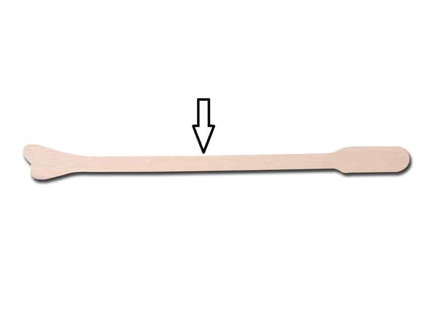
Use of Instrument marked in the Photograph is
| A |
Pap smear |
|
| B |
Cervical dilation |
|
| C |
Retraction |
|
| D |
Curetting |
Use of Instrument marked in the Photograph is
| A |
Pap smear |
|
| B |
Cervical dilation |
|
| C |
Retraction |
|
| D |
Curetting |
Papsmear (lnstrument shown: wooden Ayre’s spatula)
Ayre spatula is a device used to collect Pap smear. It is a wooden spatula with U shaped openings on one side and a flat surface on another.[1] The broad end is for vaginal sample collection and the narrow end is for cervical sample collection. It is rotated 360 degrees in the vagina to obtain the cells to be sent for Pap smear examination. Recent studies have shown that long-tipped spatulas (Aylesbury device) or a cytobrush along with an extended-tip spatula are better than Ayre spatula in collecting endocervical cells.[2] However, Ayre spatula continues to be used for cervical sample collection in less-income countries. Ayre spatula is introduced into the cervix after visualizing the os using a speculum. The cytology specimens are obtained by rotating the spatula firmly over the ectocervix and quickly transferring the cells to a slide or jar.
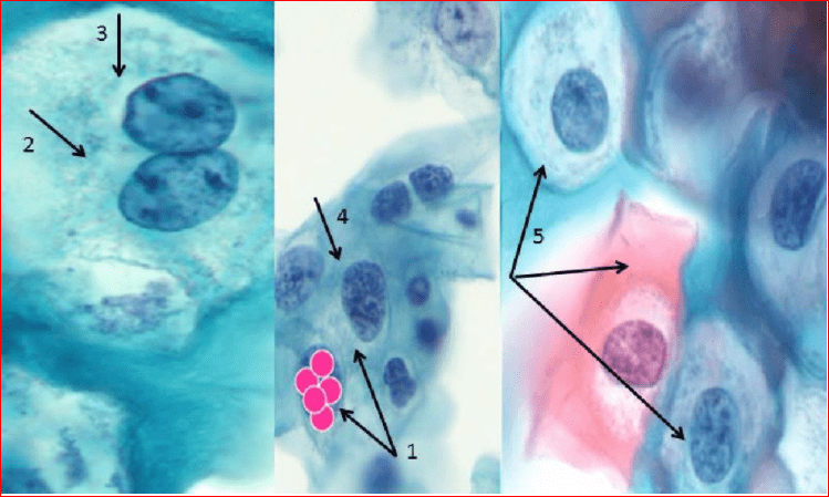
Identify this abnormality on pap smear:
| A |
Normal |
|
| B |
Adenocarcinoma |
|
| C |
Low grade Squamous Intraepithelial Lesions |
|
| D |
Squamous cell carcinoma |
Identify this abnormality on pap smear:
| A |
Normal |
|
| B |
Adenocarcinoma |
|
| C |
Low grade Squamous Intraepithelial Lesions |
|
| D |
Squamous cell carcinoma |
Ans:C.)Low grade Squamous Intraepithelial Lesions
Image shows:
1 – enlarged nuclei (approximately 5 x intermediate cell nucleus);
2 – multinucleated cells;
3 – irregular nuclear margins;
4 – presence of rough chromatin deposits;
5 – presence of koilocytes.
Pathology of squamous cells:
- Atypical Squamous Cells-Undetermined Significance(ASC-US)
- Atypical Squamous Cells – cannot exclude HSIL (ASC-H)
- Low grade Squamous Intraepithelial Lesions (LSIL)
- High grade Squamous Intraepithelial Lesions (HSIL)
- Squamous cell carcinoma
| HSIL | LSIL | |
|---|---|---|
| NC ratio | ~1:2 | ~1:3 |
| Nuclear membrane irregularities | Marked – distinct notches | Moderate |
| Chromatin granularity | Coarse, clumped, +/-nucleolus (red) | Coarse, no nucleolus |
| Cytoplasmic staining | Dark | Light |
| Perinuclear clearing | Usually absent | Often present |
| Binucleation | Uncommon | May be present |
| Maturity of squamous cell | Normal maturity | Hypermature (orangeophilic cell present) |
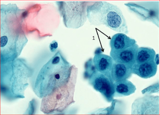
Identify this abnormality on pap smear:
| A |
Normal |
|
| B |
Squamous Cell Carcinoma |
|
| C |
Adenocarcinoma |
|
| D |
High grade Squamous Intraepithelial Lesion |
Identify this abnormality on pap smear:
| A |
Normal |
|
| B |
Squamous Cell Carcinoma |
|
| C |
Adenocarcinoma |
|
| D |
High grade Squamous Intraepithelial Lesion |
Ans:D.)High grade Squamous Intraepithelial Lesion.
Image shows:1 – increased number of parabasal cells, enlarged nuclei, irregular nuclear margins, expressed hyperchromia and the presence of rough chromatin deposits.
Pathology of squamous cells:
- Atypical Squamous Cells-Undetermined Significance(ASC-US)
- Atypical Squamous Cells – cannot exclude HSIL (ASC-H)
- Low grade Squamous Intraepithelial Lesions (LSIL)
- High grade Squamous Intraepithelial Lesions (HSIL)
- Squamous cell carcinoma.
| HSIL | LSIL | |
|---|---|---|
| NC ratio | ~1:2 | ~1:3 |
| Nuclear membrane irregularities | Marked – distinct notches | Moderate |
| Chromatin granularity | Coarse, clumped, +/-nucleolus (red) | Coarse, no nucleolus |
| Cytoplasmic staining | Dark | Light |
| Perinuclear clearing | Usually absent | Often present |
| Binucleation | Uncommon | May be present |
| Maturity of squamous cell | Normal maturity | Hypermature (orangeophilic cell present) |
Identify this abnormality on Pap Smear:

| A |
Normal |
|
| B |
Adenocarcinoma |
|
| C |
Squamous Cell Carcinoma |
|
| D |
Low Grade Squamous Intraepithelial Lesion |
Identify this abnormality on Pap Smear:
| A |
Normal |
|
| B |
Adenocarcinoma |
|
| C |
Squamous Cell Carcinoma |
|
| D |
Low Grade Squamous Intraepithelial Lesion |
Ans:C.)Squamous Cell Carcinoma.
Image shows:
1 – signs of HSIL; 2 – large nuclei and irregular chromatin deposits;3 – tumor diathesis;4 – cellular polymorphism.
Pathology of squamous cells:
- Atypical Squamous Cells-Undetermined Significance(ASC-US)
- Atypical Squamous Cells – cannot exclude HSIL (ASC-H)
- Low grade Squamous Intraepithelial Lesions (LSIL)
- High grade Squamous Intraepithelial Lesions (HSIL)
- Squamous cell carcinoma
| HSIL | LSIL | |
|---|---|---|
| NC ratio | ~1:2 | ~1:3 |
| Nuclear membrane irregularities | Marked – distinct notches | Moderate |
| Chromatin granularity | Coarse, clumped, +/-nucleolus (red) | Coarse, no nucleolus |
| Cytoplasmic staining | Dark | Light |
| Perinuclear clearing | Usually absent | Often present |
| Binucleation | Uncommon | May be present |
| Maturity of squamous cell | Normal maturity | Hypermature (orangeophilic cell present) |
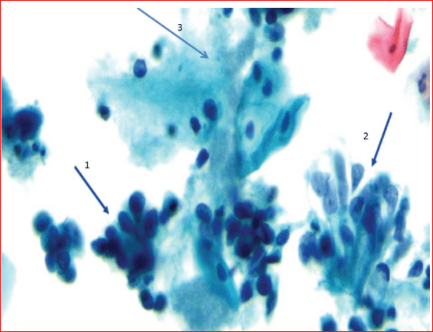
Identify this abnormality on Pap Smear:
| A |
Normal |
|
| B |
Squamous Cell Carcinoma |
|
| C |
Adenocarcinoma |
|
| D |
Adenocarcinoma in Situ |
Identify this abnormality on Pap Smear:
| A |
Normal |
|
| B |
Squamous Cell Carcinoma |
|
| C |
Adenocarcinoma |
|
| D |
Adenocarcinoma in Situ |
Ans:C.)Adenocarcinoma.
Image shows:1 – round and large nuclei; increased cellular cytoplasma;2 – macronucleoli are common;3 – tumor diathesis.
Pathology of glandular cells:
- Atypical Glandular Cells – Not Otherwise Specified (AGC-NOS) (can be classified as endocervical, endometrial or non classified) .
- Atypical Glandular Cells – Favor Neoplasia (AGC-FN) Atypical glandular cells, which are more neoplastic .
- Adenocarcinoma in situ AIS
- Adenocarcinoma
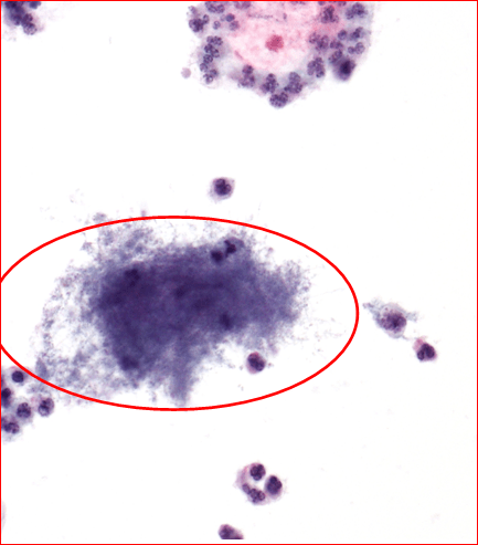
Which organsim would lead to the following abnormality in this pap smear?
| A |
HSV |
|
| B |
HPV |
|
| C |
Actinomyces |
|
| D |
Trichomonas Vaginalis |
Which organsim would lead to the following abnormality in this pap smear?
| A |
HSV |
|
| B |
HPV |
|
| C |
Actinomyces |
|
| D |
Trichomonas Vaginalis |
Ans:C.)Actinomyces.
Image shows:Clusters of Actinomyces-like organisms in the pap smear.
Actinomyces are gram positive, non acid-fact filamentous bacteria that are normally seen in cervical smears of women who have an IUCD.
The bacteria are pseudo-filamentous aggregates that can present in many shapes and sizes. They have a dusty appearance leading to it being colloquially named ‘dust bunny’
A 25 year old female patient presents with fishy odour vaginal discharge.Pap Smear shows the following picture.What can be the most possible diagnosis?

| A |
Trichomoniasis |
|
| B |
Bacterial Vaginosis |
|
| C |
Candidiasis |
|
| D |
Actinomycosis |
A 25 year old female patient presents with fishy odour vaginal discharge.Pap Smear shows the following picture.What can be the most possible diagnosis?
| A |
Trichomoniasis |
|
| B |
Bacterial Vaginosis |
|
| C |
Candidiasis |
|
| D |
Actinomycosis |
Ans:B.)Bacterial Vaginosis.
Indicator cells also known as “Clue Cells” form the characteristic picture of BV.
Note the presence of multiple microorganisms, which are attached to a single squamous epithelial cell.

