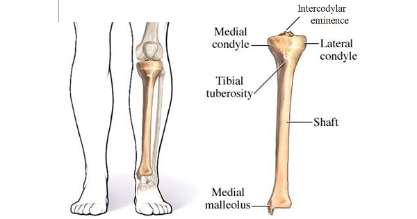
TIBIA BONE
Posterior dislocation of tibia on femur is prevented by:
| A |
Posterior cruciate ligament |
|
| B |
Anterior cruciate ligament |
|
| C |
Medial meniscus |
|
| D |
Lateral meniscus |
Posterior dislocation of tibia on femur is prevented by:
| A |
Posterior cruciate ligament |
|
| B |
Anterior cruciate ligament |
|
| C |
Medial meniscus |
|
| D |
Lateral meniscus |
A i.e. Posterior cruicate ligament
Length of tibia is:
| A |
10% of height |
|
| B |
20% of height |
|
| C |
30% of height |
|
| D |
40% of height |
Length of tibia is:
| A |
10% of height |
|
| B |
20% of height |
|
| C |
30% of height |
|
| D |
40% of height |
Ans. 20% of height
False about tibia-fibula is ‑
| A |
Nutrient artery of tibia is from posterior tibial artery |
|
| B |
Nutrient artery of fibula is from peroneal artery |
|
| C |
Proximal end of tibia is related to common peroneal nerve |
|
| D |
Tibia is the most common site of osteomyelitis |
False about tibia-fibula is ‑
| A |
Nutrient artery of tibia is from posterior tibial artery |
|
| B |
Nutrient artery of fibula is from peroneal artery |
|
| C |
Proximal end of tibia is related to common peroneal nerve |
|
| D |
Tibia is the most common site of osteomyelitis |
- Common peroneal nerve is related to neck of fibula (not tibia).
- Nutrient artery of tibia is a branch of posterior tibial artery.
- Nutrient artery of fibula is a branch of peroneal artery.
- Tibia is the commonest site of osteomyelitis.
Posterior gliding of tibia on femur is prevented by ‑
| A |
Anterior cruciate ligament |
|
| B |
Posterior cruciate ligament |
|
| C |
Medial collateral ligament |
|
| D |
Lateral collateral ligament |
Posterior gliding of tibia on femur is prevented by ‑
| A |
Anterior cruciate ligament |
|
| B |
Posterior cruciate ligament |
|
| C |
Medial collateral ligament |
|
| D |
Lateral collateral ligament |
Ans. is ‘b’ i.e., Posterior cruciate ligament
Posterior cruciate ligament
- PCL begins from posterior part of intercondylar area of tibia and runs upwards, forwards and medially to attach the anterior part of the lateral surface of medial condyle of femur.
- PCL is extrasynovial but intracapsular, i.e., lies between synovium and capsule of the knee joint.
- It provides antero-posterior stability and prevents posterior gliding of tibia on femur.
- It is taut in flexion.
- Blood supply of cruciate (anterior & posterior) ligaments is from : –
- Middle genicular artery (major supply)
- Inferior genicular (medial & lateral) artery (less important).
- Nerve supply of cruciate ligaments (ACL & PCL) is from posterior articular branch of tibial nerve.
Action of tibialis anterior ‑
| A |
Plantar flexion of foot |
|
| B |
Adduction of foot |
|
| C |
Inversion of foot |
|
| D |
None of the above |
Action of tibialis anterior ‑
| A |
Plantar flexion of foot |
|
| B |
Adduction of foot |
|
| C |
Inversion of foot |
|
| D |
None of the above |
Ans. is ‘c’ i.e., Inversion of foot
Anterolateral avulsion fracture of the distal tibial physis is known as ‑
| A |
Potts fracture |
|
| B |
Tillaux fracture |
|
| C |
Chopartracture |
|
| D |
Jones fracture |
Anterolateral avulsion fracture of the distal tibial physis is known as ‑
| A |
Potts fracture |
|
| B |
Tillaux fracture |
|
| C |
Chopartracture |
|
| D |
Jones fracture |
Ans. is ‘b’ i.e., Tillaux fracture
Tillaux fractures
- Fracture occurring in older adolescents.
- Mechanism of injury is an external rotational force with stress placed on the anterior tibio – fibular ligament, causing avulsion of the distal tibial physisanterolaterally.
- It occurs after the medial part of the physis has closed but before the lateral part closes.
- It is either Salter-Harris type III or IV fracture.
The ligaments connecting the menisci to the tibia are known as:
| A |
Coronary |
|
| B |
Arcuate |
|
| C |
Transverse |
|
| D |
Oblique |
The ligaments connecting the menisci to the tibia are known as:
| A |
Coronary |
|
| B |
Arcuate |
|
| C |
Transverse |
|
| D |
Oblique |
Ans. a. Coronary
Mechanism of injury in lateral condylar fracture of proximal tibia
| A |
Strain of valgus knee |
|
| B |
Strain of varus knee |
|
| C |
Strain of valgus knee with axial loading |
|
| D |
Rotational injury |
Mechanism of injury in lateral condylar fracture of proximal tibia
| A |
Strain of valgus knee |
|
| B |
Strain of varus knee |
|
| C |
Strain of valgus knee with axial loading |
|
| D |
Rotational injury |
Ans. is ‘c’ i.e., Strain of valgus knee with axial loading
Tibial Plateau fractures
- Mechanism of injury: Fractures of the tibial plateau are caused by yams or valgus force combined with axial loading.
- Eg: car striking a pedestrian (so called bumper fracture).
- It is more often seen in cases of fall from height in which the knee is forced into varus or valgus.
- Classification: Schatzker classification for the proximal tibia fractures.
Length of tibia is ‑
| A |
10% of height |
|
| B |
20% of height |
|
| C |
30% of height |
|
| D |
40% of height |
Length of tibia is ‑
| A |
10% of height |
|
| B |
20% of height |
|
| C |
30% of height |
|
| D |
40% of height |
Ans. is ‘b’ i.e., 20% of height
- Stature is determined in dismembered body (skeletal remains) by :
- Length from the tip of middle finger to the tip of opposite middle finger when arms are fully extended.
- Twice the length of one arm + 30 cm (of two clavicles) + 4 cm (for the sternum).
- Humerus length is 1/5th of height.
- The length from the vertex to the symphysis pubis is half of the total length.
- The length from the sternal notch to Symphysis pubis x 3-3.
- The length of forearm measured from tip of middle finger is =5/19 of total length.
- The height of head measured by the vertical distance from the top of the head (vertex) to the tip of chin = 1/8 of the total length.
- The length of vertebral column = 34/100 of total length. To the length of entire skeleton, add 2.5 to 4 cm for thickness of the soft parts.
- As a general rule humerus is 20%, tibia is 22%, femur is 27% and spine is 35% of individual height.
