
Scoliosis
Risser Localiser cast is used in the management of:
| A |
Kyphosis |
|
| B |
Spondylolysthesis |
|
| C |
Idiopathic scoliosis |
|
| D |
Lordosis |
Risser Localiser cast is used in the management of:
| A |
Kyphosis |
|
| B |
Spondylolysthesis |
|
| C |
Idiopathic scoliosis |
|
| D |
Lordosis |
Idiopathic scoliosis [Ref- Sing Med J. 1988; 29:219-221; Maheshwari Orthopedics 3/e p235]
- Risser’s Localiser cast is used for non-operative correction of idiopathic scoliosis. Other casts and braces used for scoliosis are: Milwaukee brace, Boston brace
- Operative management of idiopathic scoliosis includes:
- The Harrington Rodding
- Rod and sublaminar wiring (Luque)
- The Cotrel-Dubousset system
d.Anterior instrumentation (Dwyer; Zielke)
Casts and Splints
Minerva cast———————————— Cervical spine fracture
Hanging cast———————————— Fracture Humerus
Cylinder cast———————————– Fracture Patella
Patellar tendon bearing cast————— Fracture Tibia
Aluminium splint—————————– immobilization of finger
Thomas splint———————————- Fracture femur
Bohler Braun splint————————– Fracture femur
Toe rising splint——————————- Foot drop
Knuckle bender splint———————– Ulnar nerve palsy
Cock up splint——————————— Radial nerve palsy
Dennis Brown splint————————- CTEV
- Also Know
Risser’s sign:
This is a sign to assess skeletal maturity as the scoliotic curvature progresses most during the period of rapid skeletal growth.
The iliac apophyses start ossifying shortly after puberty. Once the iliac crests are completely ossified, further progression of the scoliosis is minimal (Risser’s sign)
| A | Fully segmented Hemivertebra | |
| B |
Wedge vertebra |
|
| C |
Block vertebra |
|
| D |
Unilateral unsegmented bar with Hemivertebra |
Block vertebra type of scoliosis has the best prognosis. The progression is slow and it can be corrected easily, whereas unilateral thoracic unsegmented type carries worst prognosis.
Ref: Orthopaedic Surgery Essentials in Pediatrics By Kathryn E. Cramer, Susan A. Scherl, 2004), Page 65; Campbell’s Orthopaedics, 11th Edition, Page 2607.
Which of the following is the MOST common skeletal manifestation in Type-1 Neurofibromatosis?
| A |
Cortical thinning of long bones |
|
| B |
Pseudoarthrosis |
|
| C |
Sphenoid dysplasia |
|
| D |
Scoliosis |
Which of the following abnormality is seen in a preoperative pulmonary function test, done in a patient with severe kyphoscoliosis?
| A |
Increased FRC |
|
| B |
Increased RV/TLC |
|
| C |
Reduced FEV1/FVC |
|
| D |
Reduced FEV25-75 |
Which type of cerebral palsy is commonly associated with scoliosis and other orthopedic problems ‑
| A |
Spastic quadriplegia |
|
| B |
Anterior cerebral palsy |
|
| C |
Spastic deplegia |
|
| D |
Atonic cerebral palsy |
Ans. is ‘a’ i.e., Spastic quadriplegia
“Scoliosis is common, occuring in 15 to 25 percent of the total body CP (spastic quadriplegia CP)”. — Tachdjian Cerebral palsy (CP)
o Cerebral palsy is defined as a non progressive neuromotor disorder of cerebral origin.
o It has following features ‑
i) Static (nonprogressive)
ii) Disorder of movement/motor disorder (but sometimes sensory involvement may occur)
iii) Cerebral in origin
Etiopathogenesis
o CP is due to an insult of developing brain.
o The insult results in maldevelopment and disorderly anatomic organization of the brain.
o The insult may be prenatal (most of the cases), during delivery or in the postnatal period.
Classification
o CP may be classified into following types –
1) Spastic cerebral palsy
2) Hypotonic (Atonic) CP
3) Extrapyramidal CP
4) Mixed type CP
- This is the most common type
- This includes
i) Spastic quadriparesis ii) Spastic diplesia iii) Spastic hemiplegia
| A |
Hypertrophy of limb |
|
| B |
Scoliosis |
|
| C |
Cafe au lait spots |
|
| D |
All |
Ans. is `d’ i.e., All
The abnormal preoperative pulmonary function test in a patient with severe kyphoscoliosis includes:
| A |
Reduced RV and TLC |
|
| B |
Reduced FEV1/FVC. |
|
| C |
Reduced FEV25-75 |
|
| D |
Increased FRC |
Answer is A (Reduced RV and TLC):
Kyphoscoliosis is a restrictive lung disease.
- There is decrease in residual volume and total lung capacity.
- FEV1 and FVC are proportionately decreased.
- Hence the ratio of FEV1 to FVC remains normal or is increased.
- The forced expiratory flow rate (FEF) is normal.
- Functional residual capacity (FRC) is decreased.
| A |
Obesity |
|
| B |
Large liver |
|
| C |
Scoliosis |
|
| D |
Congenital causes |
Ans. C.)Scoliosis.
Causes of Unilateral Elevation of Diaphragm:
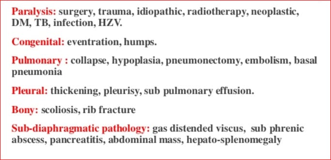
Unilateral elevation of Diaphragm occurs more commonly in left side as compared to right side.
Also,the most common cause is acquired Phrenic nerve palsy.
So,looking to the above discussion,Scoliosis may be the correct answer.
Vertebral rotation in scoliosis is checked in
| A |
Forward bending |
|
| B |
Backward bending |
|
| C |
Sideways |
|
| D |
Without bending |
Ans. is ‘a’ i.e., Forward bending
The forward bending test is very sensitive in demonstrating the vertebral rotation that takes place in a structural scoliotic curve.
- Quantification of the rotation is done by measuring the rib hump by use of inclinometer or scoliometer.
- Severity of the curve in scoliosis is measured by cobb’s angle, i.e. an angle between line passing through the margins of vertebra at ends of curve. To use the cobb method, one must first decide which vertebrae are the end-vertebrae of the curve. These end vertebrae are the vertebrae at the upper and lower limits of curve which tilt most severely toward the conavity of the curve. Other method used to measure scoliosis angle is Ferguson’s method.
| A | Scoliosis | |
| B |
Kyphosis |
|
| C |
Cubitus varus |
|
| D |
Genu varum |
Ans. is ‘a’ i.e., Scoliosis
The Milwaukee brace is principally a thoracic support consisting of a pelvic corset connected by adjustable steel supports to a cervical ring carrying occipital and chin pads.
Its purpose is to reduce the lumbar lordosis and encourage active stretching and straightening of the thoracic spine.
It is used in the conservative management of idiopathic scoliosis.
Turn-buckle cast is used for ‑
| A | Fracture shaft humerus | |
| B | Fracture shaft femur | |
| C |
Scoliosis |
|
| D |
Cervical spine injury |
Ans. is ‘c’ i.e., Scoliosis.
Name of the case Use
Minerva cast Cervical spine disease
Risser’s cast Scoliosis
Turn-buckle cast Scoliosis
Shoulder spica Shoulder immobilisation
U-slab Fracture of the humerus
Hanging cast Fracture of the humerus
Colle’s cast Colles’ fracture
Hip spica Fracture of the femur
Cylinder cast Fracture of the patella
PTB cast Fracture of the tibia
