Tuberous Sclerosis
| A |
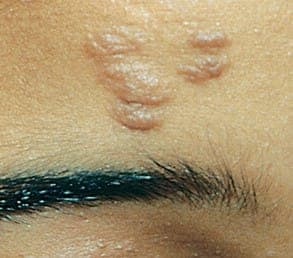
Facial angiofibroma |
|
| B |
Shagreen patch |
|
| C |
Ash leaf spot |
|
| D | Gingival fibroma |
| A |
Facial angiofibroma |
|
| B |
Shagreen patch |
|
| C |
Ash leaf spot |
|
| D | Gingival fibroma |
Ash leaf spot
REF: Harrison’s 18th ed chapter 53
“The earliest manifestation of tuberous sclerosis is Ash leaf spot”
Some form of dermatological sign will be present in 96% of individuals with TSC. Most causeno problems but are helpful in diagnosis.
Some cases may cause disfigurement, necessitating treatment. The most common skin abnormalities include:
- Facial angiofibromas (“adenoma sebaceum”): A rash of reddish spots or bumps, which appear on the nose and cheeks in a butterfly distribution. They consist of blood vessels and fibrous tissue. This socially embarrassing rash starts to appear during childhood and can be removed using dermabrasion or laser treatment. A recent publication indicates that topical rapamycin is helpful for treatment of facial angiofibromas.[71
- Periungual fibromas: Also known as Koenen’s tumors, these are small fleshy tumors that grow around and under the toenails or fingernails and may need to be surgically removed if they enlarge or cause bleeding. These are very rare in childhood but common by middle age.
- Hypomelanic macules (“ash leaf spots”): White or lighter patches of skin that may appear anywhere on the body and are caused by a lack of melanin. These are usually the only visible sign of TSC at birth. In fair-skinned individuals a Wood’s lamp (ultraviolet light) may be required to see them.
- Forehead plaques: Raised, discolored areas on the forehead.
- Shagreen patches: Areas of thick leathery skin that are dimpled like an orange peel, usually found on the lower back or nape of the neck.
Other skin features are not unique to individuals with TSC, including molluscum fibrosum or skin tags, which typically occur across the back of the neck and shoulders, cafe au Tait spots or flat brown marks, and poliosis, a tuft or patch of white hair on the scalp or eyelids.
| A | Facial angiofibroma | |
| B |
Shagreen patch |
|
| C |
Ash leaf spot |
|
| D |
Gingival fibroma |
Ash leaf spot REF: Harrison’s 18th ed chapter 53
Repeat in December 2011
“The earliest manifestation of tuberous sclerosis is Ash leaf spot”
Some form of dermatological sign will be present in 96% of individuals with TSC. Most cause no problems but are helpful in diagnosis. Some cases may cause disfigurement, necessitating treatment. The most common skin abnormalities include:
Facial angiofibromas (“adenoma sebaceum”): A rash of reddish spots or bumps, which appear on the nose and cheeks in a butterfly distribution. They consist of blood vessels and fibrous tissue. This socially embarrassing rash starts to appear during childhood and can be removed using dermabrasion or laser treatment. A recent publication indicates that topical rapamycin is helpful for treatment of facial angiofibromas.
Periungual fibromas: Also known as Koenen’s tumors, these are small fleshy tumors that grow around and under the toenails or fingernails and may need to be surgically removed if they enlarge or cause bleeding. These are very rare in childhood but common by middle age.
- Hypomelanic macules (“ash leaf spots”): White or lighter patches of skin that may appear anywhere on the body and are caused by a lack of melanin. These are usually the only visible sign of TSC at birth. In fair-skinned individuals a Wood’s lamp (ultraviolet light) may be required to see them.
- Forehead plaques: Raised, discolored areas on the forehead.
- Shagreen patches: Areas of thick leathery skin that are dimpled like an orange peel, usually found on the lower back or nape of the neck.
Other skin features are not unique to individuals with TSC, including molluscum fibrosum or skin tags, which typically occur across the back of the neck and shoulders, café au laft spots or flat brown marks, and poliosis, a tuft or patch of white hair on the scalp or eyelids.
Earliest feature of Tuberous sclerosis is:
| A | Angiofibroma | |
| B | Shagreen patch | |
| C | Ash leaf spot | |
| D | Neurofibroma |
| A |
Tuberous sclerosis |
|
| B |
Neurofibromatosis 1 |
|
| C |
Neurofibromatosis 2 |
|
| D |
All of the above |
Angiomyolipomas are present in 25% to 50% of patients with tuberous sclerosis, a disease caused by loss-of-function mutations in the TSC1 or TSC2 tumor suppressor genes.
This is a benign tumor consisting of vessels, smooth muscle, and fat.
It is characterized by lesions of the cerebral cortex that produce epilepsy and mental retardation, a variety of skin abnormalities, and unusual benign tumors at other sites, such as the heart.
| A |
Sturge Weber Syndrome |
|
| B |
Tuberous Sclerosis |
|
| C |
Neurofibromatosis |
|
| D |
Linear epidermal nevus syndrome |
A child presented to the emergency department with seizures. On examination oval hypopigmented macules were noted on the trunk, along with sub-normal IQ. What is the probable diagnosis?
| A |
Neurofibromatosis |
|
| B |
Sturge Weber |
|
| C |
Tuberous sclerosis |
|
| D |
Incontinentia Pigmenti |
Essentials of Diagnosis & Typical Features of Tuberous sclerosis:
- Facial angiofibromas or subungual fibromas.
- Often hypomelanotic macules, gingival fibromas.
- Retinal hamartomas.
- Cortical tubers or subependymal glial nodules, often calcified.
- Renal angiomyolipomas.
| A |
Facial angiofibroma |
|
| B |
Shagreen patch |
|
| C |
Ash leaf spot |
|
| D |
Gingival fibroma |
Ash leaf spots are white, ovoid, hypopigmented, ash leaf–shaped macules that can be found on the trunks or limbs. White macules offer an excellent opportunity for early diagnosis because they may be found at birth or early infancy.
Shagreen patches are flesh-colored soft plaques that are frequently found in the lumbosacral area but may occur anywhere on the trunk. The surface may be pebbly (resembling pigskin or untanned leather) with prominent follicular openings. They are usually noticed during the first decade.
The characteristic lesions are angiofibromas. These are pink or skin-colored telangiectatic papules commonly observed in the nasolabial folds and on the cheeks and chin. They usually appear in children younger than 10 years and increase in size and number until adolescence, remaining unchanged thereafter.
Other areas in which they may be observed include in and around nails (ungual fibromas), scalp, and forehead, lips, dorsa of tongue, and palate.
Triad of Tuberous Sclerosis includes all, except:
| A |
Epilepsy |
|
| B |
Low intelligence |
|
| C |
Hydrocephalus |
|
| D |
Adenoma sebaceum |
Diagnostic triad of Tuberous sclerosis includes epilepsy, mental retardation and adenoma sebaceum (facial angiofibroma).
All are manifestations of tuberous sclerosis, EXCEPT:
| A |
Retinal hamartomas |
|
| B |
Pulmonary lymphangioleiomyomatosis |
|
| C |
Posterior embryotoxon |
|
| D |
Renal angiomyolipomas |
Internal manifestations of tuberous sclerosis include seizures, mental retardation, central nervous system (CNS) and retinal hamartomas, pulmonary lymphangioleiomyomatosis (women), renal angiomyolipomas, and cardiac rhabdomyomas.
A couple has two children affected with tuberous selerosis. On detailed clinical and laboratory evaluation (including molecular studies) both parents are normal. Which one of the following explains the two affected children in this family ?
| A |
Non penetrance |
|
| B |
Uniparental diasomy |
|
| C |
Genomic imprinting |
|
| D |
Germline mosaicism |
Ans. is ‘d’ i.e. germline mosaicism
Inheritance of Tuberous sclerosis
o Tuberous sclerosis is a genetic condition which is transmitted either through inheritance or as a spontaneous genetic mutation.
o Mutation in one of the two genes TSC1 and TSC2 have been identified as a cause for tuberous sclerosis.
o Approximately 33% or one third of people with Tuberous sclerosis inherit it from a parent who also has tuberous sclerosis. This occurs via dominance inheritance.
o Thus there is 50 per cent chance with each pregnancy for a parent with Tuberous sclerosis to have a child with Tuberous sclerosis.
o In the remaining 66 % or two thirds of the people with Tuberous sclerosis, neither parent has tuberous sclerosis. o It appears that one of the normal genes from either parent changes to the abnormal form, leading to a new (or
sporadic), occurrence of tuberous sclerosis in the child. Normally these parents do not have another child with
tuberous sclerosis because the mutation was sporadic not inherited. However, some families have more than one
child with Tuberous sclerosis even though neither parent showed symptoms or findings of Tuberous sclerosis.
How does this occur?
o Scientists have determined that small number of physically unaffected parents of a child with TSC have mutations in some of their cells.
o Because the mutation is limited to a small portion of all of the body’s cells, these individuals show no signs of Tuberous sclerosis.
o But if a portion of the egg or sperm cells of a parent carries the tuberous sclerosis mutation, that parent can have
more than one affected child, possibly at the same 50/50 chance that people with Tuberous sclerosis have.
o A person who carries cells with Tuberous sclerosis mutation in her egg or his sperm supply has germline
mosaicism.
o “Mosaicism” means that the person’s body is made up of a combination of cells with and cells without a Tuberous sclerosis mutation.
o ‘Germline mosaicism’ refers to the presence of cells with Tuberous sclerosis mutations in the egg or the sperm cell supply.
o Germline mosaicism is relatively rare and this explanation does not apply to most families with a sporadically affected child. However, the occurrence of germline mosaicism has geneticists to estimate a recurrence risk (or chance that a family with a sporadically affected child will have anohter child with TSC) ranging from 1 to 3%. At this time there is no way to determine whether an unaffected parent of a child with TSC has germline mosaicism.
Type of inheritance in Tuberous sclerosis –
| A | Autosomal dominant | |
| B | Autosomal recessive | |
| C |
X-linked dominant |
|
| D |
X-linked recessive |
Ans. is ‘a’ i.e., Autosomal dominant
Drug of choice for infantile spasms in a patient with tuberous scleosis is –
| A |
Vigabatrin |
|
| B |
Tiagabine |
|
| C |
Lamotrigine |
|
| D |
Levetiracetam |
Ans. is ‘a’ i.e., Vigabatrin
A triad of seizure, mental retardation and sebaceous adenoma is seen in –
| A |
Congenital syphilis |
|
| B |
Tuberous sclerosis |
|
| C |
Toxoplasmosis |
|
| D |
Hypothyroidism |
Ans. is ‘b’ i.e., Tuberous sclerosis
Tuberous sclerosis :
o It is an autosomal dominant neurocutaneous syndrome.
o Pathology
- Characteristics brain, lesion, consists of tubers in the brain.
- They are typically present in subependyneal region where they undergo calcification producing candle dripping appearance.
| A | Neurofibromatosis type 1 | |
| B |
Tuberous sclerosis |
|
| C |
Sturge weber’s syndrome |
|
| D |
Linear Sebaceous nevus syndrome |
Ans. is ‘b’ i.e., Tuberous sclerosis
History of seizures and multiple hypopigmented macules over the back (ash leap patch) suggest the diagnosis of tuberous sclerosis.
| A |
Tuberous sclerosis |
|
| B |
Neurofibromatosis |
|
| C |
Psoriasis |
|
| D |
Alopecia aerata |
A i.e. Tuberous sclerosis
Child with h/o hypopigmented macule on back, infantile spasm and delayed milestone has
| A |
NF |
|
| B |
Sturge weber syndrome |
|
| C |
Tuberous sclerosis |
|
| D |
Nevus anemicus. |
C i.e. Tuberous sclerosis
All are seen in Tuberous sclerosis except
| A |
Iris Nodule |
|
| B |
Renal Cortical Cyst |
|
| C |
Rhabdomyoma of heart and lung |
|
| D |
Adenoma Sebaceum |
A i.e. Iris nodule
Adenoma sebaceum is a feature of:
| A |
Neurofibromatosis |
|
| B |
Tuberous sclerosis |
|
| C |
Xanthomatosis |
|
| D |
Incontinenetia pigmenti |
B i.e. Tuberous sclerosis
Babloo a 4 year male presents with history of seizures. On examination there is hypopigmented patches on face & mental retardation. Most probable diagnosis is:
| A |
Neurofibromatosis |
|
| B |
Tuberous sclerosis |
|
| C |
Sturge Weber Syndrome |
|
| D |
Incontinenta Pigmenti |
B i.e. Tuberous sclerosis
Ash leaf maculae is found in :
| A |
Tuberous sclerosis |
|
| B |
Neurofibromatosis |
|
| C |
Lymphangioma |
|
| D |
None |
A i.e. Tuberous sclerosis
Koenen’s periungual fibromas are seen in > 50% of cases with :
| A |
Tuberous sclerosis |
|
| B |
Sturge weber syndrome |
|
| C |
Alaxia telangiectasia |
|
| D |
Neurofibroatosis |
A i.e. Tuberous sclerosis
All are true regarding tuberous sclerosis except
| A |
Autosomal dominant sporadic transmission |
|
| B |
Vogt triad of epiloia |
|
| C |
Cafe au lait macules exclude the diagnosis |
|
| D |
Fibrous facial plaque |
C i.e. Cafe au lait macules exclude the diagnosis.
Tuberous sclerosis is autosomal dominant disorderQ presenting with classical Vogt’s triad of epilepsy, low intelligence/mental retardation/ delayed mile stones and adenoma sebaceum (arjgiofibroma of face)Q and ashleaf (stippled or Confetti) hypomelanotic offwhite macules mostly on trunk & buttockQ. (Acronym-Epiloia)
A child presents to the clinic with history of seizures and mental retardation. Clinical examination reveals multiple hypopigmented macules. What is the likely diagnosis:
| A |
Tuberous Sclerosis |
|
| B |
Neurofibromatosis |
|
| C |
Sturge Weber Syndrome |
|
| D |
Linear epidermal nevus syndrome |
Answer is A (Tuberous Sclerosis):
The presence of hvopigmented macules in the background of seizures and mental retardation suggests the diagnosis of Tuberous Sclerosis.
Often hypopigmented macules are the initial clue to tuberous sclerosis, a neurocutaneous disorder inherited in an autosomal dominant manner. Any child with unexplained seizures should be examined carefully for cutaneous clues, particularly hypopigmented macules. The hypopigmented lesions are small, ovoid and scattered and their number varies.’ — Oski’s Pediatrics 4th/861
The off white hypomelonotic macules are seen more easily in tanned or dark skinned individuals. The macules often are oval or ‘ash leaf’ in shape and follow dermatomes. In infancy the presence of these macules accompanied by seizures is virtually diagnostic of Tuberous Sclerosis’
Tuberous Sclerosis
Autosomal Dominant Neurocutaneous Disorder
|
Cutaneous Features |
Neurological Features |
Other Features |
|
|
• Hypopigmented macules/Ash-leaf spots° |
• Seizures° |
• Renal Lesions |
|
|
– Seen in >90% of cases |
– Seizures are the most common |
Renal cysts or angiomyolipomas |
|
|
– Are small, ovoid, scattered and variable |
presenting symptom |
may occur (Hematuria |
|
|
in number |
– Infantile spasms may be the |
/obstruction) |
|
|
– Larger lesions are known as ‘Ash-leaf’ |
presenting feature during |
• Cardiac Lesions |
|
|
spots as these may have jagged edges |
infancy |
Rhabdomyomas of the heart may |
|
|
resembling a ‘leaf |
• Mental Retardation° |
occur |
|
|
• Shagreen Spots° |
– Mental Retardation occurs in |
• Pulmonary Lesions |
|
|
– Roughened, raised, leathery lesions with an orange peel consistency • Adenoma Sebaciume – Facial skin hamartoma that develops in |
upto 50% of patients referred to tertiary care – Patients with seizures are more prone to mental retardation |
Lymphagiomyomatosis is the classical pulmonary lesion Rarely cystic lung disease may occur • Eye lesions |
|
|
The charachteristic brain lesion is a cortical ‘Tuber’. The most common neurological |
|||
|
early childhood (— 4-6 years of age) – Appears as tiny red nodules over nose |
Retinal Hamartomas may occur • Skeletal lesions |
||
|
and cheeks resembling acne • Facial Angiofibromas / Ungual Fibromas Facial angiofibromas may seen ungula fibromas are more common in toes • Café- au- lait spots |
include manifestations seizures, seizures cognitive impairment and behavioral abnormalities . including autism. |
Cystic rarefaction of the bones or the fingers or toes. |
|
|
|
|||
|
These may be seen occasionally |
|
||
|
|
|||
|
T. S. ‘Classical Triad’ described includes Seizures, Mental Retardation and Adenoma Sebacium (occurs in <33% patients) |
|||
|
T.S should be suspected in any child presenting with unexplained seizures who shows hjpopigmented cutaneous macules. |
|||
| A | Hypothyroidism | |
| B |
Tuberous sclerosis |
|
| C |
Toxoplasmosis |
|
| D |
Down syndrome |
Answer is B (Tuberous Sclerosis):
Neurological features of seizures and mental retardation together with cutaneous manifestations in the form of sebaceous adenomas are characteristic of the neurocutaneous syndrome of Tuberous sclerosis.
Triad of Tuberous Sclerosis includes all, except:
| A |
Epilepsy |
|
| B |
Adenoma sebacium |
|
| C |
Low intelligence |
|
| D |
Hydrocephalus |
The answer is D (Hydrocephalus):
The characteristic triad in tuberous sclerosis includes epilepsy, mental retardation, and Adenoma Sebacium.
Tuberous sclerosis complex (TSC) is a rare multisystem autosomal dominant genetic disease that causes non-cancerous tumors to grow in the brain and on other vital organs such as the kidneys, heart, liver, eyes, lungs, and skin.
A combination of symptoms may include seizures, intellectual disability, developmental delay, behavioral problems, skin abnormalities, lung disease, and kidney disease.
March 2007
| A | Psoriasis | |
| B |
Tuberous sclerosis |
|
| C |
Multiple sclerosis |
|
| D |
Pemphigus vulgaris |
Ans. B: Tuberous Sclerosis
Tuberous sclerosis/ tuberous sclerosis complex (TSC) is a rare, multi-system genetic disease that causes benign tumours to grow in the brain and on other vital organs such as the kidneys, heart, eyes, lungs, and skin.
TSC is caused by mutations on either of two genes, TSC1 and TSC2, which encode for the proteins hamartin and tuberin respectively.
These proteins act as tumour growth suppressors, agents that regulate cell proliferation and differentiation.
The physical manifestations of tuberous sclerosis are due to the formation of hamartia (malformed tissue such as the cortical tubers), hamartomas (benign growths such as facial angiofibroma and subependymal nodules) and, very rarely, cancerous hamartoblastomas.
Central nervous system
Classic intracranial manifestations of tuberous sclerosis include subependymal nodules and cortical/ subcortical tubers.
Kidneys
Between 60 and 80% of TSC patients have benign tumors (hamartomas) of the kidneys called angiomyolipomas (AML) frequently causing hematuria.
Lungs
Patients with TSC can develop progressive replacement of the lung parenchyma with multiple cysts. This process is identical to another disease called lymphangioleiomyomatosi (LAM).
Heart
Rhabdomyomas are benign tumors of striated muscle.
Skin
The most common skin abnormalities include:
– Facial angiofibromas (“adenoma sebaceum”): A rash of reddish spots or bumps, which appear on the nose and cheeks in a butterfly distribution.
– Ungual or subungual fibromas: Small fleshy tumors that grow around and under the toenails or fingernails.
– Hypomelanic macules (“ash leaf spots”): White or lighter patches of skin that may appear anywhere on the body and are caused by a lack of melanin.
These are usually the only visible sign of TSC at birth. In fair-skinned individuals a Wood’s lamp (ultraviolet light) may be required to see them.
– Forehead plaques: Raised, discolored areas on the forehead.
– Shagreen patches: Areas of thick leathery skin that are dimpled like an orange peel, usually found on the lower back or nape of the neck.
Eyes
Retinal lesions, called astrocytic hamartomas, appear as a greyish or yellowish-white lesion in the back of the globe on ophthalmic examination.
| A | Wegener’s granulomatosis | |
| B |
Histiocytosis-X |
|
| C |
Tuberous sclerosis |
|
| D |
Rheumatoid arthritis |
Ans. Wegener’s granulomatosis
In tuberous sclerosis all are seen except:
| A |
Subependymal nodules |
|
| B |
Tubers |
|
| C |
Giant cell astrocytoma |
|
| D |
Ependymoma |
Ans. Ependymoma
Koenen tumor seen in ‑
| A |
Neurofibromatosis |
|
| B |
Tuberous sclerosis |
|
| C |
Struge weber syndrome |
|
| D |
Tuberculosis |
Ans. is ‘b’ i.e., Tuberous sclerosis
Periungual fibromas (Koenen’s tumors) :‑
- Present in 20% of patients of tuberous sclerosis.
- Develop in adult life.
- Small, pink, sausage-shaped growths arising from under the nail folds; may distort the nail plate.
| A | Renal angiomyoma | |
| B |
Subependymal giant cell astrocytoma |
|
| C |
Rhabdomyoma heart |
|
| D |
All the above |
Ans. is ‘d’ i.e., All the above
- Tumors associated with tuberous sclerosis are subependymal giant cell astrocytoma, ependymoma, rhabdomyomas of heart, angiomyomas of kidney, liver, adrenal and pancreas.
| A |
Shagreen patch |
|
| B |
Adenoma sebacecum |
|
| C |
McCollon Plaques |
|
| D |
Depigmented nevi |
Ans. is ‘c’ i.e., McCollon Plaques
A patient presented with tuberous sclerosis on examination. The Triad of Tuberous Sclerosis includes all, except ?
| A |
Epilepsy |
|
| B |
Adenoma sebacium |
|
| C |
Low intelligence |
|
| D |
Hydrocephalus |
|
Diagnostic triad of Tuberous sclerosis includes epilepsy, mental retardation and adenoma sebaceum (facial angiofibroma).
Tuberous sclerosis (TS) is an autosomal dominant disorder which result from mutations in either the TSC1 gene encoding hamartin or the TSC2 gene encoding tuberin. Hamartin and tubulin form a complex which negatively regulate cell growth and proliferation through inhibition of mTOR.
Ref: Harrison’s Internal Medicine, 18th Edition, Chapter 284; The 5-Minute Neurology Consult By D. Joanne Lynn, Page 430; Tuberous Sclerosis Complex: Genes, Clinical Features and Therapeutics By David J. Kwiatkowsk, Page 221
|
| A | Neurofibromatosis | |
| B |
Sturge Weber |
|
| C |
Tuberous sclerosis |
|
| D |
Incontinenta pigmenti |
Ans. c. Tuberous sclerosis
- Most probable diagnosis in a child with seizures, oval hypo-pigmented macules on the trunk, and sub-normal IQ is Tuberous sclerosis.
| A |
Giant cell astrocytoma |
|
| B |
Subependymal nodule |
|
| C |
White matter lesion |
|
| D |
Ependymoma |
Ans. d. Ependymoma
Ependymoma is typically a feature of neurofibromalosis 2, but it can be seen in tuberous sclerosis as well, though it is
much less common.
Seizuresa
– Seizures are the MC presenting symptom
– Infantile spasms may be the presenting feature during infancy
Mental Retardation
Mental Retardation occurs in up to 50% of patients referred to tertiary care
Characteristic brain lesion is a cortical ‘Tuber’.
MC neurological manifestations: Seizures, cognitive impairment and behavioral abnormalities including autism.
Patients with seizures are more prone to mental retardationo
Subependymal nodules which may calcify
Hydrocephalus
Color of tuberous sclerosis lesions on wood lamp examination ‑
| A |
Bright green |
|
| B |
Milky white |
|
| C |
Golden yellow |
|
| D |
Blue white |
Ans. is ‘d’ i.e., Blue white
Wood lamp
- This is a source of ultraviolet light (320-400nm) from which virtually all visible rays have been excluded by a Wood’s (nickel oxide) filter.
- Primarily emits 360nm.
- UV light, when absorbed by certain substances, fluorescences in dark and color produced, is useful in diagnosis of the certain conditions
| Condition |
Fluorescence colour |
| Tinea capitis | Bright yellow green |
| Erythrasma |
Coral red or pink |
| Vitiligo | Milky white |
| Albinism | Blue white |
| Leprosy | Blue white |
| Tuberous sclerosis | Blue white |
| Pseudomonas infection | Greenish white |
| Porphyria | Pink/orange |
| Tinea vesicolor | Golden yellow |
Following are the differential diagnosis of congenital disorders of pigmentation ‑
| A | Teitz syndrome | |
| B | Piebaldism | |
| C |
Tuberous sclerosis |
|
| D |
All the above |
Ans. is ‘d’ i.e., All the above
Differential diagnosis of congenital disorders of pigmentation
- Waardenburg syndromes types 1-4
- Tietz syndrome
- Piebaldism
- Woolf’s syndrome
- Generalized vitiligo
- Segmental vitiligo
- Vogt-Koyanagi-Harada syndrome
- Chemical leukoderma
- Tuberous sclerosis (hypopigmented macules and patches)
- Ziprkowski-Margolis syndrome (X-linked albinism-deafness syndrome)
Koenen’s tumor associated with ‑
| A | Tuberous sclerosis | |
| B |
Neurofibromatosis |
|
| C |
Psoriasis |
|
| D |
Alopecia aerata |
Ans. is ‘a’ i.e., Tuberous sclerosis
- Koenen’s periungual fibroma is seen in tuberous sclerosis
Type of inheritance in Tuberous sclerosis ‑
| A |
Autosomal dominant |
|
| B |
Autosomal recessive |
|
| C |
X-linked dominant |
|
| D |
X-linked recessive |
Ans. is ‘a’ i.e., Autosomal dominant