Uterus
ANATOMY:


- The uterus is pyriform shape thick-walled muscular organ capable of expansion to accommodate a growing fetus.
- Measures 9 cm in length, 6.5 cm in width and 3.5 cm in thickness(3X2X1 Inches3)
- Uterus develops from Paramesonephric duct( Mullerian duct)
- It weighs 1 ounce (60 g)
- It is connected distally to the vagina, and laterally to the uterine tubes.
- The uterus has 3 parts;
- Fundus: Top of the uterus, above the entry point of the uterine tubes.
- Body: Usual site for implantation of the blastocyst.
- Cervix: Lower part of uterus linking it with the vagina. This part is structurally and functionally different to the rest of the uterus.
ANATOMICAL POSITION:
- The exact anatomical location of the uterus varies with the degree of distension of the bladder. In the normal adult uterus, it can be described as anteverted with respect to the vagina, and anteflexed with respect to the cervix:
- Anteverted: Rotated forward, towards the anterior surface of the body.
- Anteflexed: Flexed, towards the anterior surface of the body.
- Thus, the uterus normally lies immediately posterosuperior to the bladder, and anterior to the rectum.
MICROANATOMY:
- The lining epithelium of uterus is columnar
- Before menarche the cells are ciliated, but thereafter most of the cells may not have cilia Ciliated columnar epithelium
- The fundus and body of the uterus are composed of three tissue layers;
- Peritoneum – Double layered membrane, continuous with the abdominal peritoneum. Also known as the perimetrium.
- Myometrium – Thick smooth muscle layer. Cells of this layer undergo hypertrophy and hyperplasia during pregnancy in preparation to expel the fetus at birth.
- Endometrium – Inner mucous membrane lining the uterus. It can be further subdivided into 2 parts:
- Deep stratum basalis: Changes little throughout the menstrual cycle and is not shed at menstruation.
- Superficial stratum functionalis: Proliferates in response to oestrogens, and becomes secretory in response to progesterone. It is shed during menstruation and regenerates from cells in the stratum basalis layer.
LIGAMENTS:
- Pelvic diaphragm, Uterosacral ligament & Transverse cervical ligament are primary support of uterus
- The tone of the pelvic floor provides the primary support for the uterus. Some ligaments provide further support, securing the uterus in place.
They are:
- Broad Ligament: This is a double layer of peritoneum attaching the sides of the uterus to the pelvis. It acts as a mesentery for the uterus and contributes to maintaining it in position.It do not provide primary support.
CONTENT OF BROAD LIGAMENT
- Fallopian tube—upper portion
- Round ligament—anteriorly
- Ovarian ligament—posterior fold
- Vestigial structures of Wolffian body—epoophoron andparoophoron
- Vestigial structure of Wolffian duct—Gartner’s duct
- Ureter
- Uterine vessels
- Pelvic nerves
- Parametrial lymph node
- Pelvic cellular tissue condensed to form Mackenrodt’s ligament
- Infundibulopelvic ligament
- Round Ligament: A remnant of the gubernaculum extending from the uterine horns to the labia majora via the Superficial inguinal ring. It functions to maintain the anteverted position of the uterus.
- Ovarian Ligament: Joins the ovaries to the uterus.
- Cardinal Ligament: Located at the base of the broad ligament, the cardinal ligament extends from the cervix to the lateral pelvic walls. It contains the uterine artery and vein in addition to providing support to the uterus.
- Uterosacral Ligament: Extends from the cervix to the sacrum. It provides primary support to the uterus.
Supports of the genital organs:
| LEVELS | STRUCTURE |
| I |
Uterosacral ligaments and cardinal ligaments support the uterus and vaginal vault |
| II |
Pelvic facias and paracolpos which connects the vagina to the white line on the lateral pelvic wall through arcus tendinous |
| III |
Levator ani muscles support the lower one third of vagina |
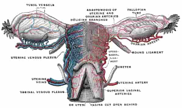
BLOOD SUPPLY AND LYMPHATICS:
- Uterine and ovarian artery
- Venous drainage is via a plexus in the broad ligament that drains into the uterine veins.
- Lymphatic drainage : iliac, sacral, Paraaortic and inguinal lymph nodes.
INNERVATION OF UTERUS:
- Sympathetic nerve fibres of the uterus arise from the uterovaginal plexus. This largely comprises the anterior and intermediate parts of the inferior hypogastric plexus.
- Parasympathetic fibres of the uterus are derived from the pelvic splanchnic nerves (S2-S4).
- The cervix is largely innervated by the inferior nerve fibres of the uterovaginal plexus.
- The afferent fibres mostly ascend through the inferior hypogastric plexus to enter the spinal cord via T10-T12 and L1 nerve fibres.
Exam Important
- Size of uterus in inches is 3x2x1
- Pelvic diaphragm, Uterosacral ligament & Transverse cervical ligament are primary support of uterus
- Paramesonephric duct forms Uterus
- Uterus develops from Mullerian duct
- External iliac , Internal iliac & Superficial inguinal recieves lymphatics from the uterus
- Uterus, before menarche, is lined by Ciliated columnar epithelium
- Blood supply of the uterus is by Ovarian & Uetrine artery
- Superficial inguinal ring in the female transmits Round ligament of the uterus
- Uterus reaches up to umbilical level at 24 weeks
- Fundus of uterus drains into Paraaortic
Don’t Forget to Solve all the previous Year Question asked on Uterus
Click Here to Start Quiz
Click Here to Start Quiz
