

Question
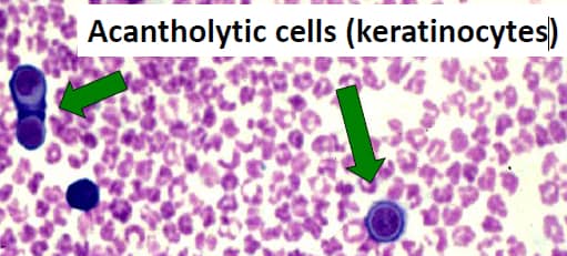
A 45 yr old female showed these cells and on direct immunofluoroscence showed intercellular IgG deposits in the epidermis. The most probable diagnosis is:

A. Bullous Pemphigoid
B. Stevens Johnson syndrome
C. Pemphigus vulgaris
D. Herpes simplex 1 infection
Show Answer


|
Correct Answer » C Explanation |
 |

This image shows Acontholytic cells in pemphigus vulgaris
Recurrent, painful oral lesions and cutaneous lesions in the form of flaccid bullae are characteristic of phemphigus vulgaris
Positive Tzanck smear, IgG deposits on direct immunofluoroscence positive nikolsky’s sign are features of phemphigus vulgaris.