Question
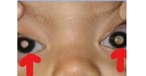
A child presents with Squint and Deterioration of Vision.On examination of the eye,following picture is seen.On ophthalmoscopic examination,a nodular cream colored mass with increased vascularization is seen.
On histopathological examination of the mass,Rossetes are seen.Regarding the radiological examination of this mass,which of the following is untrue:
A. Intraocular calcification.
B. CT scan is more sensitive for detection of extraocular extension.
C. Hetrogenous mass.
D. Necrotic components with increased vascularization.
Show Answer
|
Correct Answer » B
Explanation
|
|
Ans:B. CT scan is more sensitive for detection of extraocular extension
The child shown in the picture above is suffering from Retinoblastoma.
Retinoblastoma
- It is a cancer that rapidly develops from the immature cells of a retina.
- It is the most common malignant cancer of the eye in children, and it is almost exclusively found in young children.
- Almost half of children with retinoblastoma have a hereditary genetic defect associated with retinoblastoma.
- In other cases, it is caused by a congenital mutation in the chromosome 13 gene, 13q14.
Clinical Features:
- Abnormal appearance of the retina as viewed through the pupil(leukocoria, also known as amaurotic cat’s eye reflex).
- Deterioration of vision, a red and irritated eye with glaucoma, squint.
- Clinical patterns of retinoblastoma growth.
- Endophytic tumor,Exophytic tumor or Diffuse infiltrating growth
- Retinoblastoma can demonstrate local spread along the optic nerve directly into the orbit. It can also metastasize hematogenously to bone, liver, brain and other organs.
Examination:
- Red Reflex:Whitening of red reflex
- Ophthalmoscopic examination:
- Presents with one or multiple nodular, white or cream colored masses often associated with increased vascularization.
Histological findings:
- Both undifferentiated and differentiated elements may be present. Undifferentiated elements appear as collections of small, round cells with hyperchromatic nuclei; differentiated elements include Homer Wright rosettes(a type of rosette in which differentiated tumor cells surround the neuropil), Flexner-Wintersteiner rosettes( is a spoke and wheel shaped cell formation ),and fleurettes from photoreceptor differentiation.
Radiological imgaing:
- On imaging, they are generally characterised by a heterogeneous retinal(posterior chamber) mass with calcifications, necrotic components, and increased vascularisation on Doppler ultrasound/enhancement on CT/MRI.
- MR evaluation avoids the ionizing radiation of CT and is more sensitive for detection of extraocular extension of disease, perineural spread into the optic nerve and involvement of the subarachnoid space.