

Question
A patient presented with complains of palpitations .ECG showed the following picture.What can be the most probable diagnosis?

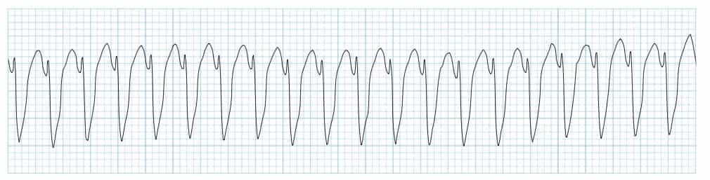
A. Monomorphic Ventricular Tachycardia.
B. Atrial Tachycardia.
C. Polymorphic Ventricular Tachycardia.
D. Atrial Fibrillation.
Show Answer


|
Correct Answer » A Explanation |
 |

Ans: A. Monomorphic Ventricular Tachycardia.
Ventricular Tachycardia (VT)
- It is a broad complex tachycardia originating in the ventricles.
Classification based on Morphology
- Monomorphic
- Polymorphic VT
- Torsades De Pointes (Polymorphic with QT prolongation)
- Right Ventricular Outflow Tract Tachycardia
- Fascicular Tachycardia
- Bidirectional VT
- Ventricular Flutter
- Ventricular Fibrillation.
ECG findings:
Features common to any broad complex tachycardia
- Rapid heart rate (> 100 bpm).
- Broad QRS complexes (> 120 ms).
Features suggestive of VT
- Very broad complexes (>160ms).
- Extreme axis deviation (“northwest axis”) — QRS is positive in aVR and negative in I + aVF.
- AV dissociation (P and QRS complexes at different rates).
- Capture beats — occur when the sinoatrial node transiently ‘captures’ the ventricles, in the midst of AV dissociation, to produce a QRS complex of normal duration.
- Fusion beats — occur when a sinus and ventricular beat coincide to produce a hybrid complex of intermediate morphology.
- Brugada’s sign – The distance from the onset of the QRS complex to the nadir of the S-wave is > 100ms.
- Josephson’s sign – Notching near the nadir of the S-wave.
- RSR’ complexes with a taller “left rabbit ear”. This is the most specific finding in favour of VT. This is in contrast to RBBB, where the right rabbit ear is taller.
Monomorphic VT
- Regular rhythm.
- Originates from a single focus within the ventricles.
- Produces uniform QRS complexes within each lead — each QRS is identical (except for usion/capture beats).