| Systematic examination of pulses |
|
|
| Which and what order? |
Where and how? |
Why? |
| 1. Radial artery |
- Radial side of wrist.
- With tips of index and middle fingers.
|
- To assess rate and rhythm.
- Simultaneously with femoral to detect delay.
- Not good for pulse character.
|
| 2. Brachial artery |
- Medial border of humerus at elbow medial to biceps tendon.
- Either with thumb of examiner’s right hand or index and middle of left hand
|
- To assess pulse character.
- To confirm rhythm.
|
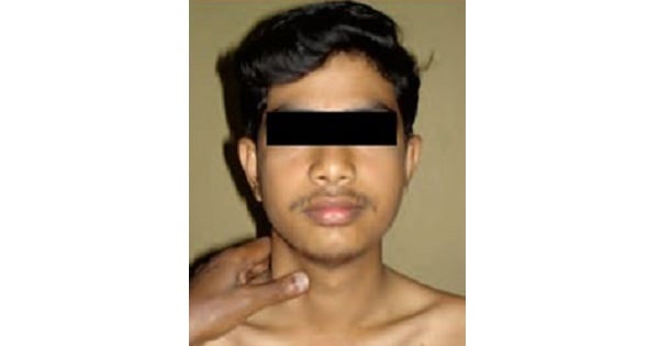
| 3. Carotid artery |
- Press examiner’s left thumb against patient’s larynx.
- Press back to feel carotid artery against precervical muscles.
- Alternatively from behind, curling fingers around side of neck.
|
- Best for pulse character and, to some extent, left ventricular function.
- To detect carotid stenosis.
- At resuscitation (CPR).
|
| 4. Femoral artery |
- Patient lying flat and undressed.
- Place finger directly above pubic ramus and midway between pubic tubercle and anterior superior iliac spine.
|
- To assess cardiac output.
- To detect radiofemoral delay.
- To assess peripheral vascular disease.
|
| 5. Popliteal artery |
- Deep within the popliteal fossa.
- Compress against posterior of distal femur with knee slightly flexed.
|
- Mainly to assess peripheral vascular disease.
- In people with diabetes.
|
| 6. Dorsalis pedis (DP) and tibialis posterior (TP) arteries (foot) |
- Lateral to extensor hallucis longus (DP).
- Posterior to medial malleolus (TP).
|
|
| 7. The abdominal aorta |
- With the flat of the hand per abdomen, as body habitus allows.
|
- In peripheral vascular disease.
- To detect aneurysmal swelling
|