Question
Identify the pathological process as shown in this histopathological examination image of Brain.
A. Coagulative Necrosis
B. Liquefactive Necrosis
C. Caseous Necrosis
D. Fat Necrosis
Show Answer
|
Correct Answer » B
Explanation
|
|
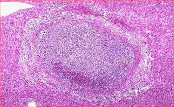
Ans:B.)Liquefactive Necrosis
Image shows:Liquefactive necrosis brain. The necrosed area on right side of the field shows a cystic space containing cell debris, while the surrounding zone shows granulation tissue and gliosis.
LIQUEFACTION (COLLIQUATIVE) NECROSIS.
- Liquefaction or colliquative necrosis occurs commonly due to ischaemic injury and bacterial or fungal infections.
- It occurs due to degradation of tissue by the action of powerful hydrolytic enzymes.
- The common examples are infarct brain and abscess cavity.
- Grossly, the affected area is soft with liquefied centre containing necrotic debris.
- Later, a cyst wall is formed.
- Microscopically, the cystic space contains necrotic cell debris and macrophages filled with phagocytosed material.
- The cyst wall is formed by proliferating capillaries, inflammatory cells, and gliosis (proliferating glial cells) in the case of brain and proliferating fibroblasts in the case of abscess cavity
Like this:
Like Loading...