Question
All the following statements are true about the cell modification marked by “C” in the image except:
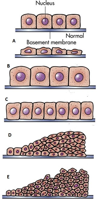
A. Increased mitoses of the cells.
B. Change of one type of cell to another type.
C. Increased rate of DNA synthesis
D. Neurons, cardiac and skeletal muscle have little or no capacity

|
Correct Answer » B Explanation |
 |
Ans:B. Change of one type of cell to another type.
Hyperplasia is shown in the image.
Change of one type of cell to another type is seen in Metaplasia.
HYPERPLASIA
- Hyperplasia is an increase in the number of parenchymal cells resulting in enlargement of the organ or tissue.
- Hyperplasia occurs due to increased recruitment of cells from G0 (resting) phase of the cell cycle to undergo mitosis, when stimulated.
- Labile cells (e.g. epithelial cells of the skin and mucous membranes, cells of the bone marrow and lymph nodes) and stable cells (e.g. parenchymal cells of the liver, pancreas, kidney, adrenal, and thyroid) can undergo hyperplasia, while permanent cells (e.g. neurons, cardiac and skeletal muscle) have little or no capacity for regenerative hyperplastic growth.
- Neoplasia differs from hyperplasia in having hyperplastic growth with loss of growth-regulatory mechanism due to change in genetic composition of the cell.
CAUSES.
A. Physiologic hyperplasia:
1. Hormonal hyperplasia e.g:
- Hyperplasia of female breast at puberty, during pregnancy and lactation.
- Hyperplasia of pregnant uterus.
- Proliferative activity of normal endometrium after a normal menstrual cycle.
- Prostatic hyperplasia in old age.
2. Compensatory hyperplasia i.e. hyperplasia occurring following removal of part of an organ or a contralateral organ in paired organ e.g.
- Regeneration of the liver following partial hepatectomy
- Regeneration of epidermis after skin abrasion
- Following nephrectomy on one side, there is hyperplasia of nephrons of the other kidney.
B. Pathologic hyperplasia. Most examples of pathologic hyperplasia are due to excessive stimulation of hormones or growth factors e.g.
- Endometrial hyperplasia following oestrogen excess.
- In wound healing, there is formation of granulation tissue due to proliferation of fibroblasts and endothelial cells.
- Formation of skin warts from hyperplasia of epidermis due to human papilloma virus.
- Pseudocarcinomatous hyperplasia of the skin.
- Intraductal epithelial hyperplasia in the breast in fibrocystic breast disease.
PATHOLOGIC FEATURES.
- There is enlargement of the affected organ or tissue and increase in the number of cells .
- This is due to increased rate of DNA synthesis and hence increased mitoses of the cells.