
Question
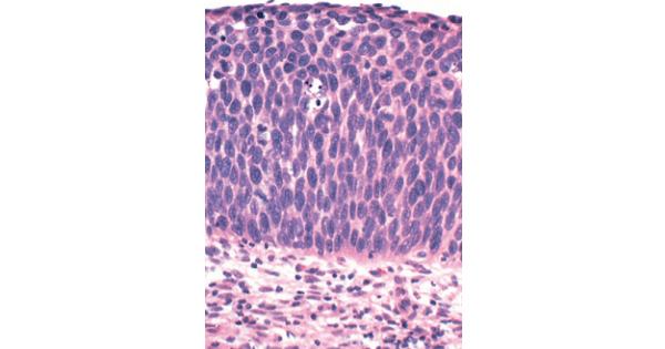
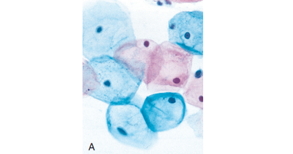
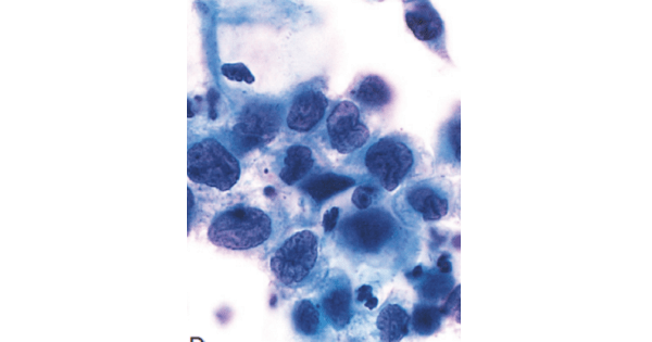
Identify the Cervical Intraepithelial Neoplasia grade by the histopathological picture and pap smear image shown.(Normal picutre is given first for comparison).

A. Stage I
B. Stage II
C. Stage III
D. Stage IV
Show Answer

|
Correct Answer » C Explanation |
 |
Ans:C. Stage III.
CERVICAL INTRAEPITHELIAL NEOPLASIA
- CIN I is characterized by dyplastic changes in the lower third of the squamous epithelium and koilocytotic change in the superficial layers of the epithelium.
- In CIN II, dysplasia extends to the middle third of the epithelium and takes the form of delayed keratinocyte maturation. It also is associated with some variation in cell and nuclear size, heterogeneity of nuclear chromatin, and presence of mitoses above the basal layer extending into the middle third of the epithelium. The superficial layer of cells shows some differentiation and occasionally demonstrates the koilocytotic changes
- The next stage, CIN III, is marked by almost complete loss of maturation, even greater variation in cell and nuclear size, chromatin heterogeneity, disorderly orientation of the cells, and normal or abnormal mitoses; these changes affect virtually all layers of the epithelium

- Spectrum of cervical intraepithelial neoplasia (CIN), with normal squamous epithelium for comparison: CIN I with koilocytotic atypia; CIN II with progressive atypia in all layers of the epithelium; and CIN III (carcinoma in situ) with diffuse atypia and loss of maturation.

- Cytologic features of cervical intraepithelial neoplasia (CIN) in a Papanicolaou smear. Superficial squamous cells may stain either red or blue. A, Normal exfoliated superficial squamous epithelial cells. B, CIN 1—low-grade squamous intraepithelial lesion (LSIL). C and D, CIN II and CIN III, respectively—both high-grade squamous intraepithelial lesions (HSILs). Note the reduction in cytoplasm and the increase in the nucleus-to-cytoplasm ratio as the grade of the lesion increases. This observation reflects the progressive loss of cellular differentiation on the surface of the cervical lesions from which these cells are exfoliated
