

CARCINOMA OF PROSTATE
CARCINOMA OF PROSTATE
- Carcinoma of prostate is the MC malignant tumour in men over 65 years.
- MC cause of bone secondaries
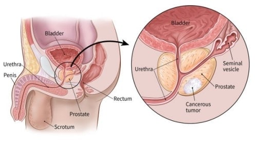
- Carcinoma of prostate occurs in peripheral zone in prostatic gland proper (commonly in posterior lobe)
RISK FACTORS-
- Advancing age + increase fat intake
- Genetic alterations is hypermethylation of glutathione transferase (GSTP-1) located on chromosome 11
PATHOLOGY-
- Adenocarcinoma of prostate is the MC form of cancer in males
- They are multifocal and heterogenous

SPREAD-
1. Local spread-
- Upward- seminal vesicles, bladder neck, trigone
- Downward- distal sphincter
2. Blood spread-
- Bones- pelvic bones, lumbar vertebrae, femoral head, ribs and skull
- MC site of origin- for skeletal metastases
3. Lymphatic spread-
- Obturator lymph nodes
CLINICAL FEATURES-
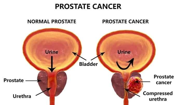
- Bladder outlet obstruction
- Haematuria
- Commonly asymptomatic
- Pelvic pain, back pain, arthritic pain
- Renal failure
- Perineural invasion
- Rectal examination- prostate feels hard, nodular, irregular and obliteration of medial sulcus
STAGING-

INVESTIGATIONS-
1. Hb%
- Anemia
- Thrombocytopenia
- DIC
2. Plain X-ray, KUB-
- Shows sclerotic metastases in lumbar vertebrae and pelvic bones
3. Serum acid phosphatase-
- Responsible for acidic pH in the prostatic urethra and normally drained in urine
- Increase acid phosphatase
4. Serum alkaline phosphatase-
- Increase in extensive liver metastasis or bone metastasis
5. Prostate specific antigen(PSA)
- It is a glycoprotein, serine protease elaborated by columnar prostatic acinar epithelial cells.
- Free- 10- 40%, complexed to antiprotease- 60 to 90%
- Formed in prostate and secreted in seminal fluid
- Causes liquefaction of seminal coagulum
- More than 4nmol/ml- carcinoma detected
- 10 nmol/ml- prostatic carcinoma
- 35 nmol/ ml- disseminated carcinoma
- Prostate specific, but not prostate cancer specific
- Most efficient test and for staging and assessing
6. Abdominal and transrectal USG- staging of the disease
7. CT scan or MRI scan
- Staging of the disease
- MRI is ideal for most accurate for local staging
8. Bone scan-
- Increased ALP
- Increased PSA (>20nmol/ml)
- For diagnosing metastasis

TREATMENT-
I) Early malignancy
a) T1 a-
- Well differentiated associated with very slow growth rate
- Regular follow up with DRE and PSA
b) T1b T1c, T2
- Radical prostactectomy or radiotherapy
- External beam radiotherapy- T1 or low T2 disease
- Branchytherapy- low T1 disease
II) Late malignancy (T3, T4 or any metastasis)
- Androgen ablation is the first line of treatment followed by antiandrogenic measure
- Orchiectomy + flutamide or LHRH + flutamide
- Palliative radiotherapy
Exam Important
- Carcinoma of prostate is the MC malignant tumour in men over 65 years.
- MC cause of bone secondaries
- Carcinoma of prostate occurs in peripheral zone in prostatic gland proper (commonly in posterior lobe)
CLINICAL FEATURES-
- Bladder outlet obstruction
- Haematuria
- Commonly asymptomatic
- Pelvic pain, back pain, arthritic pain
- Renal failure
- Perineural invasion
- Rectal examination- prostate feels hard, nodular, irregular and obliteration of medial sulcus
INVESTIGATIONS-
1. Hb%
- Anemia
- Thrombocytopenia
- DIC
2. Plain X-ray, KUB-
- Shows sclerotic metastases in lumbar vertebrae and pelvic bones
3. Serum acid phosphatase-
- Responsible for acidic pH in the prostatic urethra and normally drained in urine
- Increase acid phosphatase
4. Serum alkaline phosphatase-
- Increase in extensive liver metastasis or bone metastasis
5. Prostate specific antigen(PSA)
- It is a glycoprotein, serine protease elaborated by columnar prostatic acinar epithelial cells.
- Free- 10- 40%, complexed to antiprotease- 60 to 90%
- Formed in prostate and secreted in seminal fluid
- Causes liquefaction of seminal coagulum
- More than 4nmol/ml- carcinoma detected
- 10 nmol/ml- prostatic carcinoma
- 35 nmol/ ml- disseminated carcinoma
- Prostate specific, but not prostate cancer specific
- Most efficient test and for staging and assessing
6. Abdominal and transrectal USG- staging of the disease
7. CT scan or MRI scan
- Staging of the disease
- MRI is ideal for most accurate for local staging
8. Bone scan-
- Increased ALP
- Increased PSA (>20nmol/ml)
- For diagnosing metastasis
TREATMENT-
I) Early malignancy
a) T1 a-
- Well differentiated associated with very slow growth rate
- Regular follow up with DRE and PSA
b) T1b T1c, T2
- Radical prostactectomy or radiotherapy
- External beam radiotherapy- T1 or low T2 disease
- Branchytherapy- low T1 disease
II) Late malignancy (T3, T4 or any metastasis)
- Androgen ablation is the first line of treatment followed by antiandrogenic measure
- Orchiectomy + flutamide or LHRH + flutamide
- Palliative radiotherapy

