Fracture of the facial bones-Nose,Maxilla,Mandible,Zygomatic
FRACTURE OF THE FACIAL BONES
Fractures of the facial bones may be divided as follows:
A. Fractures of upper third-Above Supraorbital Ridge
- Frontal Sinus,Supra-orbital Ridge,Frontal bones
B. Fracture of Middle third-Between supra-orbital ridge and upper teeth
- Nasal Bones and Nasal Septum
- Naso-orbital
- Zygomatic bone
- Floor of Orbit
- Maxilla
C. Fractures of lower third
- Mandible
- Alveolus
- Temporomandibular joint
FRACTURE OF NASAL BONES
- Nasal fracture is the most common facial fracture, and the third most common fracture of the skeleton overall.
- Aetiology:Direct trauma to the nose or Face,Head injury
Types:
- Depressed-when the injury is from front
- Angulated-due to injury from side
Clinical features:
- External Nose may be swollen and deformed.Crepitus may be felt.
- Epistaxis,Nasal Blockage
Investigations:
- Radigraph of the Lateral view of the nose shows Fracture of Nasal bone with or without Displacement
- Treatment:Reduction of fracture required if the fracture fragment is displaced.
FRACTURE OF MAXILLA
It is classified into 3 types :
Le Fort I (transverse)–
- Fracture runs above and parallel to the plate. It crosses lower part of nasal septum, maxillary antra and the pterygoid plates.
Le Fort II (pyramidal) fracture–
- Passes through the root of nose, lacrimal bone, floor of orbit, upper part of maxillary sinus and pterygoid plates on both the sides.
Le Fort III (craniofacial dysjunction)–
- There is complete separation of facial bones from the cranial bones.
- The fracture line passes through root of nose, ethmofrontal junction, superior orbital fissure, lateral wall of orbit, frontozygomatic and temporozygomatic sutures and the upper part of pterygoid plates.
Clinical features:
- Depneding on the type of fracture,the following features are seen:
- Nose-Nasal bridge collapses,Epistaxis,Nasal obstruction,CSF Rhinorrhea
- CSF Rhinorrhea Occurs in fracture of maxilla in Le Fort type II and type III (as cribriform plate is injured here) and also in naso-ethmoid fracture
- Maxillary sinuses-Step deformity of the infra-orbital margin,Numbness of the cheek
- Eyes-Subconjunctival hemorrhage,Diplopia,Epiphora
- Face-Dish-face deformity with the flattening of the face
- Malocclusion of the jaws
Investigations;
- Radiograph and CT scan of Facial bones(more precise)-reveals the fractures and displacement.
Treatment
- After the Oedema of the soft tissue subsides,the fractures are reduced under general anesthesia.
- Maintainence of reduction is performed by splints,rods,bars,or continuous traction
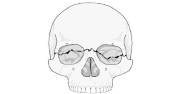
FRACTURE OF ZYGOMATIC BONE
- Zygoma fracture is also known as tripod fracture.
Clinical features :
- Considerable swelling over zygomatic arch is common
- Flattening of malar prominence.
- Step-deformity of infraorbital margin.
- Anaesthesia in the distribution of infraorbital nerve.
- Trismus, due to depression of zygoma on the underlying coronoid process.
- Oblique palpebral fissure, due to the displacement of lateral palpebral ligament.
- Restricted ocular movement, due to entrapment of inferior rectus muscle. It may cause diplopia.
- Periorbital emphysema, due to escape of air from the maxillary sinus on nose-blowing.
- The mucosa of the maxillary sinus may be lacerated and cause epistaxis on that side.
- Fracture of the zygoma may or may not be painful to palpation and running a finger along the zygomatic arch may give a feel of a depressed fracture or a small dimple.
Investigations;
- Radiograph and CT scan of Facial bones(more precise)-reveals the fractures and displacement.
Treatment
- Zygomatic arch is to be elevated.Reduction is performed by an external incision or via Caldwell -luc approach.Wiring or packing the maxillary antrum maintains the reduction of the fracture.
FRACTURE OF MANDIBLE
- Condylar process fractures of the mandible are most common account for 35% of all the fractures of mandible. They are followed by angle, body and symphysis in decreasing order of frequency.
Clinical features
- Pain,Swelling,Deformity with trismus or malocclusion of teeth
Treatment:
- The fracture is reduced and fixed by:
- Closed reduction or Open reduction.
Exam Question
- Le Forte II facial fracture implies Fracture running through zygomatic process of the maxilla, floor of orbit, root of nose on both the sides.
- Craniofacial dissociation is seen in Le Fort 3 fracture.
- CSF Rhinorrhea Occurs in fracture of maxilla in Le Fort type II and type III (as cribriform plate is injured here) and also in naso-ethmoid fracture
- Bone commonly fractured in facial injuries is Nasal Bones.
- Most common site for fracture mandible is Condyle.
- LeFort’s fracture would include Maxilla,Zygoma and Nasal Bones,
- Tripod fracture is the name given for Zygomatic fracture.
- Pyramidal fracture of maxilla is Le Fort-2
Don’t Forget to Solve all the previous Year Question asked on Fracture of the facial bones-Nose,Maxilla,Mandible,Zygomatic