Internal carotid artery
INTERNAL CAROTID ARTERY
- On angiogram internal carotid show ‘S’ shaped figure (carotid siphon).
- The internal carotid artery is a major paired artery, one on each side of the head and neck.
- They arise from the common carotid arteries where these bifurcate into the internal and external carotid arteries at cervical vertebral level 3 or 4 .
- Internal carotid artery at the bifurcation from the common carotid is Lateral to the external carotid.
- The internal carotid artery supplies the brain, while the external carotid nourishes other portions of the head, such as face, scalp, skull, and meninges.
- Supplies duramater
- Internal carotid artery lies in close relation of the optic nerve.
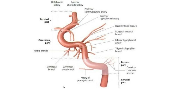
BRANCHES:
- Cervical part in the neck
- Petrous part in the petrous temporal bone
- Cavernous part in the cavernous sinus
- Cerebral part in relation to base of brain
COURSE:
1. Cervical Part:
- It ascends vertically in the neck from its origin to the base of skull to reach the lower end of the carotid canal.
- This part is enclosed in carotid sheath along with internal jugular and vagus nerve.
- No branches arises from the internal carotid artery in the neck.
- Its initial part shows slight dilation, carotid sinus, which acts as a baroreceptor.
2. Petrous Part
- Within the petrous part of the temporal bone,in the carotid canal runs upward forward & medially at rt. Angle.
Branches:
- Caroticotympanic– enter middle ear & anastomose with ant. & post. Tympanic branches
- Artery of the Pterygoid Canal– anastomose with greater palatine artery
3. Cavernous Part
- Within the Cavernous Sinus
Branches:
- Cavernous branch
- Superior & inferior Hypophyseal artery
- Meningeal branch
4. Cerebral Part-
- Lies at the base of the brain after emerging from the cavernous sinus.
- Largest
Branches:
- Ophthalmic Artery
- Anterior Cerebral Artery
- Middle Cerebral Artery
- Posterior Communicating Artery
- Anterior choroidal Artery

OPHTHALMIC ARTERY:
- Branch from cerebral part of ICA.
- It enters orbit through optic canal lying inferolateral to optic nerve.
- Both artery & nerve lie in a common dural sheath.
- It gives following branches:
1. Central artery of retina →an end artery
2. Lacrimal artery: It gives following branches-
- Lateral palpebral branch
- Zygomaticotenporal
- Zygomaticofacial
- Recurrent meningeal
- Meningeal
- Ciliary
- Anterior ethmoidal → Supplies anterior ethmoidal sinus
- Posterior ethmoidal
- Medial palpebral
- Supratrochlear
- Supraorbital
- Dorsal nasal
Exam Question
- Internal carotid artery Supplies duramater.
- Internal carotid artery lies in close relation of the optic nerve.
- Internal carotid artery at the bifurcation from the common carotid is Lateral to the external carotid
- Cavernous part of ICA runs through the medial wall of cavernous sinus.
- Internal carotid artery crosses Cavernous sinus.
BRANCHES:
1. Cervical Part:
- It ascends vertically in the neck from its origin to the base of skull to reach the lower end of the carotid canal.
- This part is enclosed in carotid sheath along with internal jugular and vagus nerve.
- No branches arises from the internal carotid artery in the neck.
- Its initial part shows slight dilation, carotid sinus, which acts as a baroreceptor.
2. Petrous Part
- Within the petrous part of the temporal bone,in the carotid canal runs upward forward & medially at rt. Angle.
- Branches:
- Caroticotympanic- enter middle ear & anastomose with ant. & post. Tympanic branches
- Artery of the Pterygoid Canal- anastomose with greater palatine artery
3. Cavernous Part
- Within the Cavernous Sinus
- Branches:
- Cavernous branch
- Superior & inferior Hypophyseal artery
- Meningeal branch
4. Cerebral Part-
- Lies at the base of the brain after emerging from the cavernous sinus.
- Largest
- Branches:
- Ophthalmic Artery
- Anterior Cerebral Artery
- Middle Cerebral Artery
- Posterior Communicating Artery
- Anterior choroidal Artery
Don’t Forget to Solve all the previous Year Question asked on Internal carotid artery