INTERNAL CAROTID ARTERY
Which is not a branch of cavernous part of internal carotid artery?
| A |
Cavernous branch |
|
| B |
Inferior hypophyseal |
|
| C |
Meningeal artery |
|
| D |
Ophthalmic artery |
Which is not a branch of cavernous part of internal carotid artery?
| A |
Cavernous branch |
|
| B |
Inferior hypophyseal |
|
| C |
Meningeal artery |
|
| D |
Ophthalmic artery |
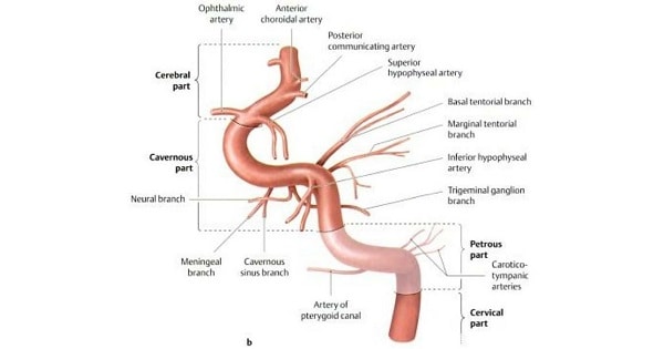
The internal carotid artery begins at the level of the upper border of the thyroid cartilage as one of the terminal branches of the common carotid artery
Its course is divided into four parts:
|
Parts of ICA |
Branches |
|
(A) Cervical part |
No branches |
|
(B) Petrous part |
• Caroticotympanic art. • Pterygoid art. |
|
(C) Cavernous part |
• Cavernous branches (to trigerninal ganglion walls of cavernous and inferior petrosal sinuses and contained nerves) |
|
|
• Sup. Hypophyseal a. |
|
|
• Inf. Hypophyseal a. |
|
|
• Meningeal a. |
|
(D) Cerebral part |
• Ophthalmic a. |
|
|
• Anterior cerebral a. |
|
|
• Middle cerebral a. |
|
|
• Post. communicating a. |
|
|
• Ant. choroidal a. |
Which of the following is not a branch of cavernous segment of Internal Carotid Artery?
| A |
Cavernous Branch |
|
| B |
Inferior Hypophyseal Branch |
|
| C |
Meningeal branch |
|
| D |
Ophthalmic branch |
Which of the following is not a branch of cavernous segment of Internal Carotid Artery?
| A |
Cavernous Branch |
|
| B |
Inferior Hypophyseal Branch |
|
| C |
Meningeal branch |
|
| D |
Ophthalmic branch |
The ophthalmic artery is a branch of the cerebral part of the internal carotid artery.
All of the following are branches of cerebral part of the internal carotid artery, EXCEPT?
| A |
Ophthalmic artery |
|
| B |
Anterior cerebral artery |
|
| C |
Posterior communicating artery |
|
| D |
Meningeal artery |
All of the following are branches of cerebral part of the internal carotid artery, EXCEPT?
| A |
Ophthalmic artery |
|
| B |
Anterior cerebral artery |
|
| C |
Posterior communicating artery |
|
| D |
Meningeal artery |
Meningeal artery is a branch of cavernous part of Internal carotid artery.
Must know:
The internal carotid artery begins at the superior border of the thyroid cartilage as one of the terminal branches of common carotid artery.
| Parts of ICA | Branches |
| (A) Cervical parts | No branches |
| (B) Petrous part |
|
| (C) Cavernous part |
|
| (D) Cerebral part |
|
The internal carotid artery has four segments: the cervical, the intrapetrosal, the cavernous, and the cerebral. Which is NOT a branch of cavernous part of internal carotid artery?
| A |
Cavernous branch |
|
| B |
Inferior hypophyseal artery |
|
| C |
Meningeal artery |
|
| D |
Ophthalmic artery |
The internal carotid artery has four segments: the cervical, the intrapetrosal, the cavernous, and the cerebral. Which is NOT a branch of cavernous part of internal carotid artery?
| A |
Cavernous branch |
|
| B |
Inferior hypophyseal artery |
|
| C |
Meningeal artery |
|
| D |
Ophthalmic artery |
The cavernous part of the internal carotid artery gives rise to numerous small branches:
- Cavernous branches: Supply the trigeminal ganglion, the walls of the cavernous, and inferior petrosal sinuses, and the nerves contained.
- A minute meningeal branch: Supply dura mater and bone in the anterior cranial fossa.
- Numerous small hypophysial branches: Supply the neurohypophysis to form the pituitary portal system.
TRUE about posterior communicating artery is ?
| A |
A branch of internal carotid artery |
|
| B |
A branch of superior cerebral artery |
|
| C |
A branch of middle cerebral artery |
|
| D |
Supplies crus cerebri |
TRUE about posterior communicating artery is ?
| A |
A branch of internal carotid artery |
|
| B |
A branch of superior cerebral artery |
|
| C |
A branch of middle cerebral artery |
|
| D |
Supplies crus cerebri |
- Anterior cerebral artery (left and right)
- Anterior communicating artery
- Internal carotid artery (left and right)
- Posterior cerebral artery (left and right)
- Posterior communicating artery (left and right)
Anterior choroidal artery is a branch of which of the following arteries?
| A |
Basilar artery |
|
| B |
Anterior cerebral artery |
|
| C |
Posterior cerebral artery |
|
| D |
Internal carotid artery |
Anterior choroidal artery is a branch of which of the following arteries?
| A |
Basilar artery |
|
| B |
Anterior cerebral artery |
|
| C |
Posterior cerebral artery |
|
| D |
Internal carotid artery |
- Meningohypophyseal artery
- Inferolateral artery
- Capsular artery
- Ophthalmic artery
- Superior hypophyseal artery
- Posterior communicating artery
- Anterior choroidal artery
- Anterior cerebral artery
- Middle cerebral artery
All of the following are the branches of intracavernous part of the internal carotid artery except:
| A |
Hypophysial branches |
|
| B |
Ophthalmic artery |
|
| C |
Branch to trigemical ganglion |
|
| D |
Meningeal artery |
All of the following are the branches of intracavernous part of the internal carotid artery except:
| A |
Hypophysial branches |
|
| B |
Ophthalmic artery |
|
| C |
Branch to trigemical ganglion |
|
| D |
Meningeal artery |
B i.e., Opthalmic artery
No. of branches of the internal carotid artery in the neck is:
| A |
1 |
|
| B |
2 |
|
| C |
3 |
|
| D |
None |
No. of branches of the internal carotid artery in the neck is:
| A |
1 |
|
| B |
2 |
|
| C |
3 |
|
| D |
None |
D i.e. None
Ophthalmic artery is the branch of….part of internal carotid artery :
| A |
Intercavernous |
|
| B |
Intrapetrous |
|
| C |
Intracerebral |
|
| D |
Extra cranial |
Ophthalmic artery is the branch of….part of internal carotid artery :
| A |
Intercavernous |
|
| B |
Intrapetrous |
|
| C |
Intracerebral |
|
| D |
Extra cranial |
C- i.e. Intracerebral
Internal carotid artery at the bifurcantion from the common carotid is –
| A |
Lateral to the extermal carotid |
|
| B |
Medial to external carotid |
|
| C |
Post to external carotid |
|
| D |
Anterior to external carotid |
Internal carotid artery at the bifurcantion from the common carotid is –
| A |
Lateral to the extermal carotid |
|
| B |
Medial to external carotid |
|
| C |
Post to external carotid |
|
| D |
Anterior to external carotid |
A i.e. Lateral to the external carotid artery
What is the nearest relation of the optic nerve
| A |
Pituitary Stalk |
|
| B |
Internal Carotid Artery |
|
| C |
Anterior Choroidal Artery |
|
| D |
Anterior Communicating Artery |
What is the nearest relation of the optic nerve
| A |
Pituitary Stalk |
|
| B |
Internal Carotid Artery |
|
| C |
Anterior Choroidal Artery |
|
| D |
Anterior Communicating Artery |
B. i.e. Internal corotid artery
What is not true for facial artery :
| A |
Main source of oxygentaed blood to palatine tonsil |
|
| B |
Is a branch of internal carotid artery |
|
| C |
Supplies branches to both upper and lower lips |
|
| D |
Conveys postganglionic sympathetic nerve fibres |
What is not true for facial artery :
| A |
Main source of oxygentaed blood to palatine tonsil |
|
| B |
Is a branch of internal carotid artery |
|
| C |
Supplies branches to both upper and lower lips |
|
| D |
Conveys postganglionic sympathetic nerve fibres |
B i.e. Is a branch of internal carotid artery
Investigation of choice for screening of proximal internal carotid artery stenosis is :
| A |
Doppler flow USG |
|
| B |
CT substraction angiography |
|
| C |
MRI |
|
| D |
Angiography (DSA) |
Investigation of choice for screening of proximal internal carotid artery stenosis is :
| A |
Doppler flow USG |
|
| B |
CT substraction angiography |
|
| C |
MRI |
|
| D |
Angiography (DSA) |
Answer is A (Doppler flow USG):
‘Stenosis at the origin of the internal carotid Artery can be identified and quantified reliably by ultrasonography that combines B mode ultrasound image with a Doppler ultrasound assessment of flow velocity.’
Arterial supply to dura mater is from all of the following except
| A |
Middle meningeal artery |
|
| B |
Internal carotid artery |
|
| C |
Ascending pharyngeal artery |
|
| D |
Basilar artery |
Arterial supply to dura mater is from all of the following except
| A |
Middle meningeal artery |
|
| B |
Internal carotid artery |
|
| C |
Ascending pharyngeal artery |
|
| D |
Basilar artery |
- Numerous arteries supply the dura mater from the internal carotid, maxillary, ascending pharyngeal, occipital, and vertebral arteries.
- From a clinical standpoint, the most important is the middle meningeal artery, which is commonly damaged in head injuries.
- The middle meningeal artery arises from the maxillary artery in the infratemporal fossa. To enter the cranial cavity, it passes through the foramen spinosum to lie between the meningeal and endosteal layers of dura.
- The anterior (frontal) branch’s course corresponds roughly to the line of the underlying precentral gyrus of the brain. The posterior (parietal) branch curves backward and supplies the posterior part of the dura mater.
Angiographically, the typical “beaded” or “pile of plates” appearance involving the internal carotid artery is observed in:
| A |
Takayu’s disease |
|
| B |
Non-specific aorto-arteritis |
|
| C |
Fibromuscular dysplasia |
|
| D |
Rendu-Osler-Weber Disease |
Angiographically, the typical “beaded” or “pile of plates” appearance involving the internal carotid artery is observed in:
| A |
Takayu’s disease |
|
| B |
Non-specific aorto-arteritis |
|
| C |
Fibromuscular dysplasia |
|
| D |
Rendu-Osler-Weber Disease |
Ans. Fibromuscular dysplasia
