
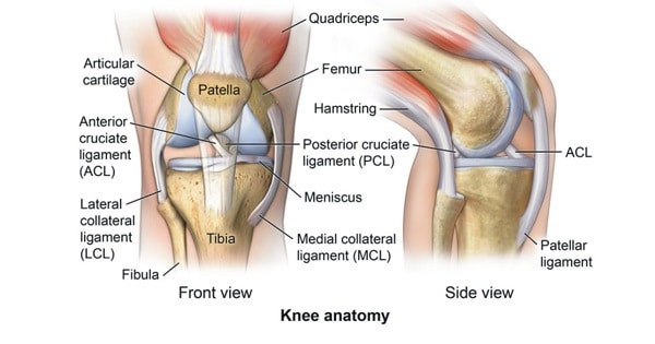
Ligaments of knee joint
LIGAMENTS OF KNEE JOINT
- The ligaments surrounding the knee joint offer stability by limiting movements and, together with the menisci and several bursae, protect the articular capsule.
INTRACAPSULAR LIGAMENT:
- The knee is stabilized by a pair of cruciate ligaments.
- The anterior cruciate ligament (ACL)
- One of the most important ligament of the knee joint.
- The ACL is critically important because it prevents the tibia from being pushed too far anterior relative to the femur.
- Begins from anterior part of intercondylar area of tibia just behind the anterior horn of medial meniscus.
- Provide proprioceptive function of knee joint.
- ACL is intrasynovial i.e it lies inside the synovium.
- ACL is taut during extension.
- Middle genicular artery is a branch of popliteal artery and supplies the cruciate ligaments & the synovial membrane of knee joint piercing the oblique popliteal ligament of knee.
2. The posterior cruciate ligament (PCL)
- Originates from Posterior part of intercondylar area of tibia.
- It stretches from medial condyle of femur to the posterior intercondylar area.
- Injury to this ligament is uncommon but can occur as a direct result of forced trauma to the ligament.
- This ligament prevents posterior displacement of the tibia relative to the femur.
- It is taut in flexion.
- PCL is extrasynovial but intracapsular i.e lies b/w synovium & capsule of the knee joint.
Blood supply of anterior & posterior ligaments is from:
- Middle genicular artery (major supply)
- Inferior genicular artery (medial & lateral)
- Nerve supply of cruciate ligaments (ACL & PCL) is from posterior articular branch of tibial nerve.
3. The transverse ligament
- It stretches from the lateral meniscus to the medial meniscus.
- It passes in front of the menisci.
- It is divided into several strips in 10% of cases.
- The two menisci are attached to each other anteriorly by the ligament.
4. The posterior and anterior meniscofemoral ligaments
- Stretch from the posterior horn of the lateral meniscus to the medial femoral condyle.
- They pass posteriorly behind the posterior cruciate ligament.
- The posterior meniscofemoral ligament is more commonly present (30%); both ligaments are present less often.
5. Coronary Ligament
- Part of capsule that connects the peripheral part of menisci with corresponding tibial condyles.
 EXTRACAPSULAR LIGAMENTS:
- Ligamentum patella (patellar tendon)
- It connects the patella to the tuberosity of the tibia.
- occasionally called the patellar tendon because there is no definite separation between the quadriceps tendon (which surrounds the patella) and the area connecting the patella to the tibia.
- This very strong ligament helps give the patella its mechanical leverage and also functions as a cap for the condyles of the femur.
2. Tibial collateral (medial collateral) ligament
- It stretches from the medial epicondyle of the femur to the medial tibial condyle.
- It is composed of three groups of fibers, one stretching between the two bones, and two fused with the medial meniscus.
- The MCL is partly covered by the pes anserinus and the tendon of the semimembranosus passes under it.
- MCL injury is most common ligament injury to the knee.
- Phylogenetically this ligament injury is the most common ligament injury to the knee.
3. Fibular (lateral) collateral ligament
- It stretches from the lateral epicondyle of the femur to the head of fibula.
- It is separate from both the joint capsule and the lateral meniscus.
- It protects the lateral side from an inside bending force (a varus force).
- The anterolateral ligament (ALL) is situated in front of the LCL.
- In comparison to deep part of tibial collateral ligament it is not attached to capsule & lateral meniscus.
4. Oblique popliteal ligament
- An expansion from the tendon of semimembranous attachment to intercondylar line of femur.
- Closely related to popliteal artery & is pierced by middle genicular vessels & nerve & the terminal part of the posterior division of the obturator nerve.
5. Arcuate popliteal ligament
- It is a posterior expansion from the short lateral ligament.
- Extends from the head of the fibula to the posterior border of the tibial intercondylar area.
Exam Question
- Middle genicular artery is a branch of popliteal artery and supplies the cruciate ligaments & the synovial membrane of knee joint
- Posterior cruciate ligament Prevents posterior dislocation of tibia
- Anterior cruciate ligament prevents Anterior dislocation of tibia
- Middle genicular artery pierces the oblique popliteal ligament of knee
- Posterior cruciate ligament Originates from Posterior part of intercondylar area of tibia
Don’t Forget to Solve all the previous Year Question asked on Ligaments of knee joint
