
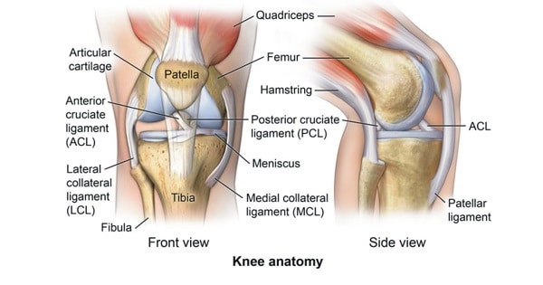
LIGAMENTS OF KNEE JOINT
| A | Fibular head artery | |
| B | Descending genicular artery | |
| C | Superior genicular artery | |
| D | Middle genicular artery |
| A | Fibular head artery | |
| B | Descending genicular artery | |
| C | Superior genicular artery | |
| D | Middle genicular artery |
Middle genicular artery
-Middle genicular artery is a branch of popliteal artery and supplies the cruciate ligaments & the synovial
membrane of knee joint.
-lt reaches the interior of knee by piercing the oblique popliteal ligament of the knee.
-Fibular head artery (A) is the circumflex fibular branch given by the posterior tibial artery, which contributes towards the anastomosis around the knee joint.
-Descending genicular artery (B) is a branch of femoral artery and also contributes to the knee joint anastomosis.
-Superior genicular artery (C) is a branch of popliteal artery and is an important branch in the anastomosis around the knee joint.
All of the following maintain the stability of ankle joint except:
| A |
Cruciate ligament |
|
| B |
Shape of the bones |
|
| C |
Tendons of muscle which cross the joint |
|
| D |
Collateral ligament |
All of the following maintain the stability of ankle joint except:
| A |
Cruciate ligament |
|
| B |
Shape of the bones |
|
| C |
Tendons of muscle which cross the joint |
|
| D |
Collateral ligament |
Cruciate ligament
Cruciate ligaments are found in the knee and not ankle. Cruciate ligaments are pairs of ligaments arranged like a letter X. They are the anterior cruciate ligament (ACL) and the posterior cruciate ligament (PCL). They maintain anteroposterior stability of the knee joint.
Which of the following is true about posterior cruciate ligament?
| A |
Attached to lateral femoral condyle |
|
| B |
Intrasynovial |
|
| C |
Prevents posterior dislocation of tibia |
|
| D |
Relaxed in full flexion |
Which of the following is true about posterior cruciate ligament?
| A |
Attached to lateral femoral condyle |
|
| B |
Intrasynovial |
|
| C |
Prevents posterior dislocation of tibia |
|
| D |
Relaxed in full flexion |
Posterior cruciate ligament is stronger than the ACL and attaches to the posterior intercondylar area of the tibia. The posterior cruciate ligament ascends in an anteromedial direction to attach on the lateral surface of the medial femoral condyle.The posterior cruciate ligament resists posterior translation of the tibia on the femur or anterior translation of the femur on the tibia.
Posterior cruciate ligament prevents the following action of the tibia on femur?
| A |
Anterior dislocation |
|
| B |
Posterior dislocation |
|
| C |
Rotation |
|
| D |
Limit hyperflexion |
Posterior cruciate ligament prevents the following action of the tibia on femur?
| A |
Anterior dislocation |
|
| B |
Posterior dislocation |
|
| C |
Rotation |
|
| D |
Limit hyperflexion |
Functions of posterior cruciate ligament:
- Stabilises knee
- Prevents posterior dislocation of the tibia on femur
- Limits hyper extension of knee only if the anterior cruciate is ruptured
Anterior cruciate ligament prevents :
| A |
Anterior dislocation of tibia |
|
| B |
Posterior dislocation of tibia |
|
| C |
Anterior dislocation of femur |
|
| D |
Posterior dislocation of femur |
Anterior cruciate ligament prevents :
| A |
Anterior dislocation of tibia |
|
| B |
Posterior dislocation of tibia |
|
| C |
Anterior dislocation of femur |
|
| D |
Posterior dislocation of femur |
A i.e. Anterior dislocation of tibia
Posterior dislocation of tibia on femur is prevented by:
| A |
Posterior cruciate ligament |
|
| B |
Anterior cruciate ligament |
|
| C |
Medial meniscus |
|
| D |
Lateral meniscus |
Posterior dislocation of tibia on femur is prevented by:
| A |
Posterior cruciate ligament |
|
| B |
Anterior cruciate ligament |
|
| C |
Medial meniscus |
|
| D |
Lateral meniscus |
A i.e. Posterior cruicate ligament
Artery piercing the oblique popliteal ligament of knee –
| A |
Superior genicular |
|
| B |
Inferior genicular |
|
| C |
Middle genicular |
|
| D |
Popliteal |
Artery piercing the oblique popliteal ligament of knee –
| A |
Superior genicular |
|
| B |
Inferior genicular |
|
| C |
Middle genicular |
|
| D |
Popliteal |
Middle genicular
- Oblique popliteal ligament is an expansion from the tendon of semimembranosus attachment to intercondylar line of femur.
- It is closely related to popliteal artery and is pierced by middle genicular vessels and nerve and the terminal part of the posterior division of the obturator nerve.
Posterior gliding of tibia on femur is prevented by ‑
| A |
Anterior cruciate ligament |
|
| B |
Posterior cruciate ligament |
|
| C |
Medial collateral ligament |
|
| D |
Lateral collateral ligament |
Posterior gliding of tibia on femur is prevented by ‑
| A |
Anterior cruciate ligament |
|
| B |
Posterior cruciate ligament |
|
| C |
Medial collateral ligament |
|
| D |
Lateral collateral ligament |
Ans. is ‘b’ i.e., Posterior cruciate ligament
Posterior cruciate ligament
- PCL begins from posterior part of intercondylar area of tibia and runs upwards, forwards and medially to attach the anterior part of the lateral surface of medial condyle of femur.
- PCL is extrasynovial but intracapsular, i.e., lies between synovium and capsule of the knee joint.
- It provides antero-posterior stability and prevents posterior gliding of tibia on femur.
- It is taut in flexion.
- Blood supply of cruciate (anterior & posterior) ligaments is from : –
- Middle genicular artery (major supply)
- Inferior genicular (medial & lateral) artery (less important).
- Nerve supply of cruciate ligaments (ACL & PCL) is from posterior articular branch of tibial nerve.
Origin of PCL- Posterior cruciate ligament
| A |
Posterior part of intercondylar area of tibia |
|
| B |
Anterior part of intercondylar area of tibia |
|
| C |
Medial part of medial femoral condyle |
|
| D |
Lateral part of medial femoral condyle |
Origin of PCL- Posterior cruciate ligament
| A |
Posterior part of intercondylar area of tibia |
|
| B |
Anterior part of intercondylar area of tibia |
|
| C |
Medial part of medial femoral condyle |
|
| D |
Lateral part of medial femoral condyle |
Posterior cruciate ligament (PCL)
- PC L begins from posterior part of intercondylar area of tibia and runs upwards, forwards and medially to attach the anterior part of the lateral surface of medial condyle of femur.
- PCL is extrasynovial but intracapsular, i.e. lies between synovium and capsule of the knee joint.
- It provides antero-posterior stability and prevents posterior gliding of tibia on femur.
- It is taut in flexion.
- Blood supply of cruciate (anterior & posterior) ligaments is from :-
- Middle genicular artery (major supply).
- Inferior genicular (medial & lateral) artery (less important).
- Nerve supply of cruciate ligaments (ACL & PCL) is from posterior articular branch of tibial nerve.

