Ligaments of larynx & epiglottis
EPIGLOTTIS
- It is an unpaired cartilage of larynx.
- Oblong leaf shaped in adult & omega shaped & larger in infants
- It develops from 4th arch
- Located behind the root of the tongue and the body of the hyoid bone and in front of the laryngeal entrance (laryngeal aditus or vestibule).
- It has 2 ends— upper & Lower
- 2 surfaces— Anterior & Posterior
- 2 Lateral borders:
- Upper end: broad
- Lower end: narrow –“ petiolus / stalk ” attaches to inner surface of thyroid cartilage below thyroid notch by the thyroepiglottic ligament
- It attaches to the posterior body of the hyoid bone via the hyoepiglottic ligament
- Epiglottis has a lingual and laryngeal surface.
- Epithelial lining of lingual surface of epiglottis is Stratified squamous epithelium
- It lies dorsal to the thyroid cartilage and thyrohyoid membrane, guarding the laryngeal entrance.
- The space between the anterior surface of the epiglottis and the thyrohyoid membrane and thyroid cartilage is called the preglottic space
- Suprahyoid epiglottis with False cords- Arytenoids forms epilarynx
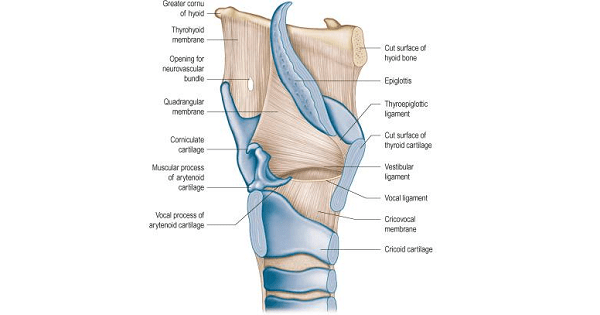
- The epiglottis is attached to the thyroid cartilage by the thyroepiglottic ligament.
- Epiglottis lies separating the esophagus from the larynx
- The aryepiglottic folds and the quadrangular membranes attach to the lower part of the lateral margins of the epiglottis.
- Most common site of sarcoidosis in larynx.
LIGAMENT & FOLDS
- Epiglottic ligaments
- Aryepiglottic fold
- Vestibular ligament (vestibular folds or false vocal cords)
- Vocal ligaments
Epiglottic Ligaments and Folds
- Hyoepiglottic ligament
- Thyroepiglottic ligament
- Median glossoepiglottic ligament
- Lateral glossoepiglottic or pharyngoepiglottic fold,
- Attached between the base of the epiglottic cartilage and the pharyngeal wall at the root of the tongue
Aryepiglottic Folds
- One on each side, forms inlet of larynx
- Contain the aryepiglottic muscles.
- Associated with the superior border of the quadrangular membrane.
- Both aryepiglottic folds constrict the entrance to the larynx and protect the respiratory pathway by not permitting food, liquids, and foreign bodies to enter the larynx and trachea.
Vestibular Folds (False Vocal Cords)
- Formed by the inferior edge of the quadrangular membrane.
- Attached in front to the thyroid cartilage just below the attachment of the epiglottic cartilage
- Connected behind to the anterolateral surfaces of the arytenoid cartilages.
- The vestibular ligaments are located just above the vocal ligaments, separated from them by bilateral ellipsoid spaces called the laryngeal ventricles.
- Overlap the true vocal folds just prior to a cough or sneeze — reinforcing the resistance offered by the true vocal folds against the internal expiratory pressures.
Vocal Ligaments, Vocal Cords, and Vocal Folds
- The thickened, ligamentous, upper edges of the elastic tissue of the conus are the vocal ligaments or vocal cords.
- Extend from the medial extremities of the laminae of the thyroid cartilage in the midline anteriorly (forming the anterior commissure) to the apices of the vocal processes of the arytenoid cartilages on each side posteriorly.
Structure of Vocal cord
- Histologically 5 layers:
- LAYER 1: is the Stratified squamous non-keratinized epithelium. It is very thin and helps to hold the shape of the vocal fold. This layer doesnot contain any mucous glands.
- LAYER 2: superfical layer of the lamina propria. It is composed of loose fibers and matrix .This layer contains only minimal elastic and collagenous fibers and offers least resistance to vibration. The integrity of this layer is vital for proper phonatory function.
- LAYER 3: intermediate layer of lamina propria.It contains a higher concentration of elastic and collagenous fibers when compared to layer 2. This layer is thickened at the anterior and posterior ends of the vocal folds. These thickened regions are known as anterior and posterior macula flava. These structures provide protection to the vocal folds from mechanical damage.
- LAYER 4 : deep layer of lamina propria.It contains a dense collection of elastic and collagenous fibers. This layer along with the intermediate layer constitute the vocal ligament. Some of the collagenous fibers present here gets inserted into the vocalis muscle.
- LAYER 5: formed by the vocalis muscle. The fibers of this muscle run parallel to the direction of the vocal fold.
- Vocalis muscle is infact a portion of thyro arytenoid muscle act as tensor of vocal cord.
- At the anterior most portion of the vocal fold a mass of collagenous tissue is present known as the anterior commissure tendon or Broyle’s ligament.
- This ligament gets attached to the inner area of thyroid cartilage which is devoid of perichondrium.
- Lacking a submucosa and blood vessels, the vocal ligaments appear to be pearly white and shiny.
- The space between the true vocal cords (the intermembranous space) is known as the rima glottidis
- RIMA GLOTTIDIS Subdivided into 2 parts,
- 2/5 – intercartilaginous part (respiratory glottis,or interarytenoid space), between the arytenoid cartilages and
- 3/5 — the intermembranous part or glottis vocalis.
Exam Question
- Epiglottis is omega shaped in infants
- Epiglottis is common site of sarcoidosis in larynx
- Epiglottis lies separating the esophagus from the larynx
- Suprahyoid epiglottis with False cords- Arytenoids forms epilarynx
- Inlet of larynx is formed by aryepiglottis fold
- Epiglottis develops from 4th arch
- Epithelial lining of lingual surface of epiglottis is Stratified squamous epithelium
- Epiglottis is an unpaired cartilage of larynx
- Stratified squamous non-keratinized epithelium is Epithelial lining of glottis/ true vocal cords
Don’t Forget to Solve all the previous Year Question asked on Ligaments of larynx & epiglottis