
OCCULOMOTOR (III) NERVE
OCCULOMOTOR NERVE
- This is purely motor (somatic motor) nerve.
STRUCTURE:
Midbrain
↓
Third nerve nucleus( at the level of the superior colliculus ventral to the cerebral aqueduct, on the pre-aqueductal grey matter)
↓
Red Nucleus
↓
Substantia Nigra
↓
Exit through Interpeduncular fossa
↓
Invested with a sheath of pia mater
↓
Passes between the superior cerebellar & posterior cerebral arteries (Nerve compressed by aneurysm of posterior communicating artery)
↓
Pierces the dura mater
↓
Pass b/w free and attached borders of tentorium cerebelli
↓
Cavernous sinus
↓
Receives filaments from the cavernous plexus of the sympathetic nervous system and communicating branch from V1
↓
Superior orbital fissure
↓
Orbit
↓
Superior and Inferior Branch
BRANCHES OF OCCULOMOTOR NERVE:
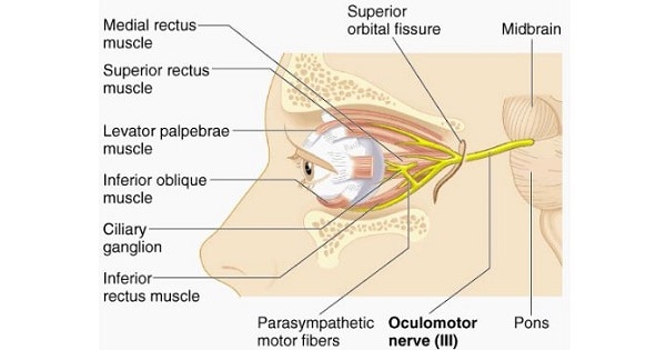
- Occulomotor nerve descends to the lateral wall of cavernous sinus and in the anterior part of sinus it divides, into upper (superior) and lower (inferior) divisions, which enter the orbit through middle part of superior orbital fissure.
1) Superior Branch:
- The superior branch of the oculomotor nerve or the superior division, the smaller, passes medially over the optic nerve.
- It supplies the superior rectus and levator palpebrae superioris.
2) Inferior Branch:
- The inferior branch of the oculomotor nerve or the inferior division, the larger, divides into three branches.
- One passes beneath the optic nerve to the medial rectus.
- Another, to the inferior rectus.
- The third and longest runs forward between the inferior recti and lateralis to the inferior oblique.
- Nerve to inferior oblique is the longest of these and gives of parasympathetic root to ciliary ganglion before supplying the muscle.
- All branches enter extraocular muscles and their ocular surfaces except for inferior oblique which enters its posterior border.
NUCLEI:
1. General somatic efferent:
- Through oculomotor nucleus for movement of eyeball supplying all extraocular muscles except Superior Oblique (SO) and Lateral Rectus (LR).
2. General visceral efferent (parasymapthetic):
- Through Edinger- Westphal nucleus for pupillary contraction and accomodation.
3. General somatic afferent:
- Carries proprioceptive fibres from the extraocular muscles to mesencephalic nucleus of trigeminal.
Occulomotor nucleus (for general somatic efferent) and Edinger- Westphal nucleus together form oculomotor nuclear complex.
FEATURES OF OCCULOMOTOR (3rd) NERVE PALSY:
1. Paralysis of superior rectus, inferior rectus, medial rectus and inferior oblique.
- Eye is down and out due to unopposed action of superior oblique and lateral rectus.
- Medial & upward gaze is not possible.
2. Paralysis of sphincter pupillae→ Loss of ipsilateral direct and consensual pupillary light reflex and there is mydriasis.
3. Paralysis of ciliary muscle→ Paralysis of accomodation and conversion reaction.
4. Two extraocular muscles are spared-
- Superior oblique: Intorsion, abduction and depression remain active.
- Lateral rectus: Abduction remains active. So eye is deviated laterally.
5. Paralysis of levator palpebrae superioris→ Ptosis.
6. Oculomotor nerve is the most commonly involved cranial nerve in intracranial aneurysm.
Exam Important
- Nerve compressed by aneurysm of posterior communicating artery is occulomotor nerve.
- The muscles innervated by occulomotor nerve are superior rectus, levator palpebrae superioris ,medial rectus, inferior rectus & inferior oblique.
- Occulomotor nerve Carries parasympathetic nerve fibres to eye.
- Causes constriction of pupil is caused by occulomotor nerve.
- Cross section of midbrain at the level of superior colliculus shows 3rd CN & red nucleus.

