
Thalassemia
INTRODUCTION:
- Most common type of hemoglobinopathies.
- An Autosomal recessive condition.
- Caused by inherited mutations decreasing synthesis of either α-globin or β-globin chains of HB.
- Two α chains in HbA encoded by alpha globin genes on chromosome 16.
- Two β chains are encoded by β-globin gene on chromosome 11.
- Diminished synthesis leads to,
- Low intracellular hemoglobin (hypochromia).
- Effects related to relative excess of the other available chain.
α-THALASSEMIAS
- Usually caused by deletions of one or more of 4 alpha globin genes.
- Presents with hepatosplenomegaly & edema.
- Basophilic stippling is absent.
- Electrophoresis in alpha-thalassemia trait – Shows normal HbF & normal HbA2.
|
Hydrops fetalis |
Tetramers of excess γ globin chains (Barts Hb) Very high affinity for O2
|
|
Haemoglobin H disease Most common in Asians |
Tetramers of excess β globin (HbH) High affinity for O2. Resemble thalassemia intermedia with moderately severe hemolytic anemia |
|
α-Thalassemia-1 trait |
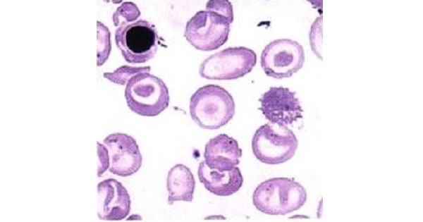
Similar to β-thalassemia minor Asymptomatic Acanthocytes (cells with irregularly spaced spiked projections) Reticulocyte count and iron parameters are normal. |
|
α Thalassemia-2 trait |
No red cell abnormality |
β THALASSEMIAS
- Caused by point mutations affecting synthesis of beta-globin chains.
Types of mutations:
- β0 mutations: Due to Splicing mutations (most commonly) & also, promoter region mutations resulting in total absence of β-globin synthesis.
- β+ mutations: Due to chain terminator mutations resulting in reduced (but detectable) β-globin synthesis.
- ↓ synthesis of structurally normal β chains coupled with increased production of α chains
- Imbalance between α- and β-globin synthesis results in unpaired alpha chains precipitation within red cell precursors → membrane damage → apoptosis & ineffective erythropoiesis.
THALASSEMIA MAJOR:
- Also referred to as“Cooley’s anaemia” or Mediterranean anaemia
Clinical features
- Anemia manifests 6 – 9 months after birth as Hb synthesis switches from HbF to HbA
Results in,
- Ineffective erythropoiesis
- Increased absorption of dietary iron & secondary hemochromatosis/hemosiderosis.
- Extramedullary hematopoiesis
- Frontal bossing, Prominent facial bones (chipmunk facies) and dental malocclusion
- Hepatosplenomegaly
Lab features
- MCV, MCHC, MCH and TIBC decreased.
- Osmotic fragility decreased.
- Haptoglobin levels decreased.
- Increased serum iron, ferritin & % transferrin saturation.
- Bone marrow – Hypercellular with reversed myeloid: erythroid ratio.
- Target cells (Characteristic finding).
- Referred as “Codocytes” – Pathognomic of beta thalassemia major.
- These are RBC’s with lump of hemoglobinized cytoplasm within area of normal central pallor resulting in “Bull’s eye” target.
- Basophilic stippling – Constant finding.
- These are ribosomal inclusions in RBCs.
- Microcytic hypochromic anemia.
- Anisocytosis (Marked variation in size) & poikilocytosis (marked variation in shape of blood cells).
- Nucleated red cells (normoblasts).

Investigations:
- Hb electrophoresis – Gold standard investigation
- Increased HbF (30% – 90%).
X-ray –
- Crew haircut appearance
- Hair on end appearance of skull bone.

NESTROFT (Naked Eye Single Tube Red cell Osmotic Fragility Test):
- Used for screening.
- Blood mixed with 0.35% normal saline.
Interpretation:
- Black-line – Normal & occurs due to hemolysis.
- No black line – Indicates Thalassemia case since RBC are resistant to lysis.

- Test for 13- thalassemia trait is HbA2
Treatment
- Severely transfusion dependent.
- Bone marrow transplantation – Only curative therapy
- Oral desferoxamine – Used for chelation therapy.
β Thalassemia intermedia
- Chronic intracorpuscular hemolytic anemia.
- Haemoglobin levels usually >7mg/dl.
β Thalassemia minor or Thalassemia trait(β+/β or β0/β)
- Asymptomatic, mild or no anaemia.
- No need for transfusions.
- Resistance against falciparum malaria.
- Peripheral smear shows – Microcytic hypochromic anemia, basophilic stripping & target cells.
Lab findings:
- Increased HbA2 – Diagnostic of beta thalasemmia minor
|
Features |
Normal |
Thalassemia minor |
Thalassemia intermedia |
Thalassemia major |
|
HbA |
97 – 99% |
80 – 95 % |
0 – 30% |
0 – 10% |
|
HbA2 |
1 – 3 % |
4 – 8 % |
0-10 |
4 – 10 % |
|
HbF |
< 1% |
1-5% |
6-100% |
90 – 96 % |
- Oral deferiprone is the most appropriate drug used for chelation therapy in beta-thalassemia major.
- Splenomegaly, target cells on peripheral smear, microcytic hypochromic anaemia are seen in thalassemia major.
- Hb-A2 estimation will be diagnostically helpful in a case of beta thalassemia trait.
- Hb-H disease is a form of alpha thalassemia.
- Intracorpuscular hemolytic anemia is seen in Thalassemia
- Defect leading to thalassemia lies in Haemoglobin.
- Thalassemia occurs due to Splicing mutation.
- In Beta thalassemia, there is decrease in beta chain & increase in alpha chain.
- A2 concentration in thalassemia trait is >3.5
- The investigation done to establish the diagnosis of thalassemia is Hb-electrophoresis.
- NESTROFT test is used in screening of Thalassemia.
- In α-thalassemia, there is no α-chain
- Thalassemia gives protection against malaria.
- Bone marrow iron is increased in Thalassemia.
- Decrease in osmotic fragility cause hemolysis in Thalassemia.
- Test for beta-thalassemia trait is HbA2
- Hair on end appearance is seen in X-ray skull in Thalassemia.
- Hepatosplenomegaly and edema all over body of new born leading to death is diagnosed to have α-thalassemia
- Splenectomy is least useful in Thalasemia major
- Normal levels of free Erythrocyte Protoporphyrin with hypochromic microcytic anemia, seen in Thalassemia
- Haptaglobin levels are decreased in Thalasemia
- Thalassemia is inherited as Autosomal recessive
- In alpha-thalassemia trait,electrophoresis shows Normal HbF and normal HbA2
- Defect in Snurps causes thalassemia
