
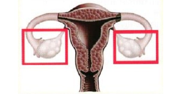
Anatomy Of Ovaries
Ovary develop from :
| A |
Mullerian duct |
|
| B |
Genital ridge |
|
| C |
Genital tubercle |
|
| D |
Mesonephric duct |
Ovary develop from :
| A |
Mullerian duct |
|
| B |
Genital ridge |
|
| C |
Genital tubercle |
|
| D |
Mesonephric duct |
Genital ridge
Friends, I am giving you a very important tabls. Many questions can be solvod trom lhis very table.
Male and female derivatives of embryonic urogenital structures.
| Embryonic structures | Derivatives | |
| Male | Female | |
| Labioscrotal swelling | Scrotumo | Labia majorao |
| Urogenilal folds | Ventral aspect ot penis | Labia minora |
| Geniial tubercleo | Peniso | Clitoriso |
| Urogenital sinus | Urinary bladder | Urinary bladder |
| Urethra except navicular lossa | Urethral and paraurethral glands | |
| Prostate gland | Vagina | |
| Prostatic utricle | Bartholin’s glands | |
| Bulbo-uretheral glands | ||
| ParamesoneDhric duct | Appendix of tesies | Hvdatid ol moroaoni. uterus. cewix |
| fallopian tubes | ||
| Mesonephric duct | Ductus eoididvmis | Duct of epoophoron |
| Garlner’s cvsl | ||
| Seminal vesiclos | ||
| Mesonephric tubules | Ductuli etterentes | Epoophoron |
| Paradidymis | Paraoophoron | |
| Undifferenliated gonads/ | Testis | Ovary |
| Genital / gonadal ridge | ||
| Cortex | Seminiferous tubules | Ovarian tollicle |
| Medulla | Rete testis | Rete ovarii |
| Gubernaculum | Gubernaculum testis | Ovarian ligament |
| Round ligament | ||
Also know :
- Germ cells are of endodermal origin and migrate from yolk sac to genital ridge between 20 – 30 days.°
- Until the seventh week of development gonads do not develop male or female characteristic (i.e. are bipotential in nature).°
- SRY gene located on the short arm of Y chromosome, directly control the development of testis from the bipotential gonads.°
- In a XX individual, without the active influence of Y chromosome, the bipotential gonad develops into an ovary two weeks later than testicular development.°
In a study it is observed that the right ovary ovulates more than left, all are possible explanations for the cause except-
| A |
Anatomical asymmetry |
|
| B |
Difference in blood supply to both sides |
|
| C |
Right handedness is more common in population |
|
| D |
Some embryological basis |
In a study it is observed that the right ovary ovulates more than left, all are possible explanations for the cause except-
| A |
Anatomical asymmetry |
|
| B |
Difference in blood supply to both sides |
|
| C |
Right handedness is more common in population |
|
| D |
Some embryological basis |
Right handedness is more common in population [Ref: huinrep.oxforcijournal.org/content/12/8/]730full.pdf]
- In the primate it is suggested that ovulations occur with equal .frequency in the left and right ovary.
- In the humans it there is some controversy about the frequency of ovulation on each side.
- It is believed that in normally menstruating women ovulation was significantly higher in right ovary.
- It is believed that right sided predominance was either genetically determined or due to difference in the vasculature of the ovaries.
- The anatomical asymmetry between the left and right side are also thought to be the reason.
- The left ovarian vein drains to the left renal vein and the right ovarian vein to the inferior venacava.
- The left renal vein is thought to be under high pressure than the right and therefore drain slower. Because the left ovary drains slower, the collapsed follicle (corpus luteum) takes longer to clean and thereby diminishes the chances that ovulation will occur on that side the .following month.
– No such condition exists on the right side which is why successive right side ovulation is more common.
The main portion of the ovarian artery, vein, and nerves are carried to and from the ovary by way of the:
| A |
Suspensory ligament |
|
| B |
Ovarian ligament |
|
| C |
Broad ligament |
|
| D |
Mesovarium |
The main portion of the ovarian artery, vein, and nerves are carried to and from the ovary by way of the:
| A |
Suspensory ligament |
|
| B |
Ovarian ligament |
|
| C |
Broad ligament |
|
| D |
Mesovarium |
The mesovarium, the posterior extension of the broad ligament, supports the ovary and becomes continuous with the serosa (or germinal epithelium) of the ovary.
Ovary is supplied by which nerve root?
| A |
T9 |
|
| B |
T10 |
|
| C |
L4 |
|
| D |
T1 |
Ovary is supplied by which nerve root?
| A |
T9 |
|
| B |
T10 |
|
| C |
L4 |
|
| D |
T1 |
The ovaries are paired organs situated close to the wall on either side of the pelvis minor, a little below the brim.
Each measures 2.5–5 cm in length, 1.5–3 cm in breadth, and 0.7–1.5 cm in width, weighing about 4–8 g.
The nerve supply of the ovaries arises from T10.
The ovary is suspended by means of the mesovarium, the suspensory ligament of the ovary, and the ovarian ligament.
Cells found in the interstitium of the ovary that resemble steroid secreting cells and may secrete androgens in the human ovary are:
| A |
Ovarian hilar cells |
|
| B |
Corpus albicans |
|
| C |
Corona radiata cells |
|
| D |
Granulosa lutein cells |
Cells found in the interstitium of the ovary that resemble steroid secreting cells and may secrete androgens in the human ovary are:
| A |
Ovarian hilar cells |
|
| B |
Corpus albicans |
|
| C |
Corona radiata cells |
|
| D |
Granulosa lutein cells |
The corpus albicans is formed after degeneration of the corpus luteum, in which the granulosa lutein cells undergo autolysis and a hyaline material accumulates within the cellular remnants of the former corpus luteum. It will eventually blend into the stromal tissue of the ovary. The ovarian hilar cells are cells found in the hilum of the ovary and may be androgen secreting cells.
Formation of primary ovary in female foetus takes place by
| A |
6th week |
|
| B |
8th week |
|
| C |
10th week |
|
| D |
12th week |
Formation of primary ovary in female foetus takes place by
| A |
6th week |
|
| B |
8th week |
|
| C |
10th week |
|
| D |
12th week |
C i.e. 10 weeks
Germ cells in the ovary develop from ‑
| A |
Coelomic endoderm |
|
| B |
Trophoblastic layer |
|
| C |
Yolk sac |
|
| D |
Surface ectoderm |
Germ cells in the ovary develop from ‑
| A |
Coelomic endoderm |
|
| B |
Trophoblastic layer |
|
| C |
Yolk sac |
|
| D |
Surface ectoderm |
C i.e. Yolk Sac
- Umbilical vesicle (Yolk sac) attains full development & is large at 32 days (4-5 weeks). It can be observed sonographically early in 5th week. By 10 weeks, it shrinks to pear shaped 5 mm diameter and after 20 weeks it is not visible.
- Although umbilical vesicle contains no yolk, its essential due to
– Site of origin of primordial germ cells is endodermal lining of wall of umbilical vesicle (in 3rd week)
– In 3rd week blood first develop in vascularized extraembryonic mesoderm covering the wall of umbilical vesicle, and continues to form there until hemopoietic activity begins in liver during 6th week
Its endoderm, derived from epiblast, gives rise to epithelium of trachea, bronchi, lungs and digestive tract (form primordial gut in 4th week)
Transfer nutrients to embryo in 2nd & 3rd weeks
Lymphatic drainage of ovary is through:
| A |
Preaortic and para-aortic lymph nodes |
|
| B |
Superficial inguinal lymph nodes |
|
| C |
Deep inguinal lymph nodes |
|
| D |
Internal iliac lymph nodes |
Lymphatic drainage of ovary is through:
| A |
Preaortic and para-aortic lymph nodes |
|
| B |
Superficial inguinal lymph nodes |
|
| C |
Deep inguinal lymph nodes |
|
| D |
Internal iliac lymph nodes |
Lymph Drainage of ovary
The lymph vessels of the ovary follow the ovarian artery and drain into the pre aortic and para-aortic nodes at the level of the first lumbar vertebra.
Arterial Supply
The ovarian artery arises from the abdominal aorta at the level of the first lumbar vertebra.
Venous drainage.
The ovarian vein drains into the inferior vena cava on the right side and into the left renal vein on the left side. Nerve Supply
The nerve supply to the ovary is derived from the aortic plexus and accompanies the ovarian artery.
Not true about development of ovary ‑
| A |
Develops in genital ridge |
|
| B |
Sex cords are derived from coelomic epithelium |
|
| C |
Oocytes are mesodermal in origin |
|
| D |
At birth ovary contains 2 million follicles |
Not true about development of ovary ‑
| A |
Develops in genital ridge |
|
| B |
Sex cords are derived from coelomic epithelium |
|
| C |
Oocytes are mesodermal in origin |
|
| D |
At birth ovary contains 2 million follicles |
Development of ovary
- Coelomic epithelium on medial side of the mesonephros becomes thickened to form genital ridge, the site where ovary develops.
- Genital ridge is covered by germinal epithelium (previous coelomic epithelium). From these germinal epithelium, cords of cells (sex cords or medullary cords) proliferate and grow into the underlying mesoderm.
- Primordial germ cells which are developed from endodermal cells of hindgut (part of yolk sac), migrate to region of developing ovary (genital ridge area) and give rise to oocytes.
- The sex cords become broken up into small masses. The cells of each mass surround one oocyte to form primordial follicle.
- At birth each ovary contains about 2 million primary follicles.
Ovarian fossa is formed by all except‑
| A |
Obliterated umbilical artery |
|
| B |
Internal iliac artery |
|
| C |
Ureter |
|
| D |
Round ligament of ovary |
Ovarian fossa is formed by all except‑
| A |
Obliterated umbilical artery |
|
| B |
Internal iliac artery |
|
| C |
Ureter |
|
| D |
Round ligament of ovary |
Each ovary lies in ovarian fossa on lateral pelvic wall which is bounded :‑
- Anteriorly: Obliterated umbilical artery
- Posteriorly: Ureter and internal iliac artery
Posterior border of ovary is ‑
| A |
Attached to suspensory ligament |
|
| B |
Attached to broad ligament |
|
| C |
Attached to infundibulopelvic ligament |
|
| D |
Free of ligament attachment |
Posterior border of ovary is ‑
| A |
Attached to suspensory ligament |
|
| B |
Attached to broad ligament |
|
| C |
Attached to infundibulopelvic ligament |
|
| D |
Free of ligament attachment |
Ans. is ‘d’ i.e., Free of ligament attachment
OVARY
The ovaries are female gonads. Each ovary lies in the ovarian fossa on the lateral pelvic wall which is bound anteriorly by obliterated umblical artery and posteriorly by ureter and internal iliac artery. Each ovary has two poles (upper and lower), two borders (anterior and posterior) and two surfaces (medial and lateral).
Upper pole (tubal end) is related to ovarian fimbria of fallopian tube and external iliac artery. Suspensory ligament of ovary (infundibulopelvic ligament) connents it with pelvic brim.
Lower pole (uterine end) is connected with lateral angle of uterus by ligament of the ovary, just posteroinferior to the attachment of fallopian tube.
Lateral surface is related to ovarian fossa and is covered with peritoneum which separates it from Obturator vessels and nerve.
Medial surface is related to fallopian tube and is separated by a peritoneal recess, called ovarian bursa.
Anterior border (mesovarian border) is straight and related to fallopian tube and obliterated umblical artery. It is attached to posterior surface of broad ligament of uterus by mesovarium and forms the hilus of ovary.
Posterior border (free border) is convex and is related to fallopian tube and ureter.
Primordial Germ cell of ovary are developed from ‑
| A |
Ectoder |
|
| B |
Mesoderm |
|
| C |
Endoderm |
|
| D |
Coelomic epithelium |
Primordial Germ cell of ovary are developed from ‑
| A |
Ectoder |
|
| B |
Mesoderm |
|
| C |
Endoderm |
|
| D |
Coelomic epithelium |
Ans. is ‘c’ i.e., Endoderm
Which among the following are not true about development of an organ marked in the picture below ? 
| A |
Develops in genital ridge. |
|
| B |
Sex cords are derived from coelomic epithelium. |
|
| C |
Oocytes are mesodermal in origin. |
|
| D |
At birth ovary contains 2 million follicles. |
Which among the following are not true about development of an organ marked in the picture below ?
| A |
Develops in genital ridge. |
|
| B |
Sex cords are derived from coelomic epithelium. |
|
| C |
Oocytes are mesodermal in origin. |
|
| D |
At birth ovary contains 2 million follicles. |
The organ marked in the picture above represents ovary.
Development of ovary
- Coelomic epithelium on medial side of the mesonephros becomes thickened to form genital ridge, the site where ovary develops.
- Genital ridge is covered by germinal epithelium (previous coelomic epithelium). From these germinal epithelium, cords of cells (sex cords or medullary cords) proliferate and grow into the underlying mesoderm.
- Primordial germ cells which are developed from endodermal cells of hindgut (part of yolk sac), migrate to region of developing ovary (genital ridge area) and give rise to oocytes.
- The sex cords become broken up into small masses. The cells of each mass surround one oocyte to form primordial follicle.
- At birth each ovary contains about 2 million primary follicles.
Number of primodial follicles in ovary at birth are‑
| A |
2 million |
|
| B |
6 million |
|
| C |
5 million |
|
| D |
10 million |
Number of primodial follicles in ovary at birth are‑
| A |
2 million |
|
| B |
6 million |
|
| C |
5 million |
|
| D |
10 million |
Ans. is ‘a’ i.e., 2 million
- In humans, no new ova are formed after birth. During fetal development, the ovaries contain over 7 million primordial follicles.
- However, many undergo atresia (involution) before birth and others are lost after birth.
- At the time of birth, there are 2 million ova, but 50% of these are atretic.
- The million that are normal undergo the first part of the first meiotic division at about this time and enter a stage of arrest in prophase in which those that survive persist until adulthood.
- Atresia continues during development, and the number of ova in both of the ovaries at the time of puberty is less than 300,000.
- Only one of these ova per cycle (or about 500 in the course of a normal reproductive life) normally reaches maturity; the remainder degenerate.

