
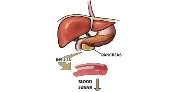
Diabetes Ketoacidosis
High Anion Gap Acidosis is seen in all EXCEPT:
| A | Diabetic Ketoacidosis | |
| B |
Lactic Acidosis |
|
| C | Renal Tubular Acidosis | |
| D |
Methanol Poisoniong |
High Anion Gap Acidosis is seen in all EXCEPT:
| A | Diabetic Ketoacidosis | |
| B |
Lactic Acidosis |
|
| C | Renal Tubular Acidosis | |
| D |
Methanol Poisoniong |
Renal Tubular Acidosis REF: Harrison’s Internal Medicine 17′ edition chapter 48 Table 48-4, http://en.wikipedia.org/ wiki/Anion_gap#High anion_ gap
Repeat from June 2009
All evaluations of acid-base disorders should include a simple calculation of the AG; it represents those unmeasured anions in plasma (normally 10 to 12 mmol/L) and is calculated as follows: AG = Na’ – (C1- + HCO3-). When acid anions such as acetoacetate and lactate accumulate in extracellular fluid, the AG increases causing a high AG acidosis. An increase in the AG is most often due to an increase in unmeasured anions and less commonly is due to a decrease in unmeasured cations (calcium, magnesium, potassium).
CAUSES OF HIGH-ANION-GAP METABOLIC ACIDOSIS:
|
Lactic acidosis |
Toxins |
|
Ketoacidosis |
• Ethylene glycol |
|
• Diabetic |
• Methanol |
|
• Alcoholic |
• Salicylates |
|
• Starvation |
• Propylene glycol |
|
• Pyroglutamic acid |
|
|
|
Renal failure (acute and chronic) |
| A | Diabetic Ketoacidosis | |
| B |
Lactic Acidosis |
|
| C | Renal Tubular Acidosis | |
| D |
Methanol Poisoning |
| A | Diabetic Ketoacidosis | |
| B |
Lactic Acidosis |
|
| C | Renal Tubular Acidosis | |
| D |
Methanol Poisoning |
Renal Tub2ular Acidosis REF: Harrison’s Internal Medicine 17th edition chapter 48 Table 48-4, http://en.wikipedia.org/ wiki/Anion_gap#High anion_gap
Repeat from December 2010
All evaluations of acid-base disorders should include a simple calculation of the AG; it represents those unmeasured anions in plasma (normally 10 to 12 mmol/L) and is calculated as follows: AG = Na’ – (C1- + HCO3-). When acid anions such as acetoacetate and lactate accumulate in extracellular fluid, the AG increases causing a high AG acidosis. An increase in the AG is most often due to an increase in unmeasured anions and less commonly is due to a decrease in unmeasured cations (calcium, magnesium, potassium).
CAUSES OF HIGH-ANION-GAP METABOLIC ACIDOSIS
Lactic acidosis Toxins
Ketoacidosis Ethylene glycol
Diabetic Methanol
Alcoholic Salicylates
Starvation Propylene glycol Pyroglutamic acid Renal failure (acute and chronic)
Rajesh is known to have IDDM and he is brought to the emergency room in a coma due to diabetic ketoacidosis. Insulin therapy is begun without a delay. Which of the following should also accompany immediately?
| A |
Calcium supplementation |
|
| B |
Creatinine supplementation |
|
| C |
Magnesium supplementation |
|
| D |
Potassium supplementation |
Rajesh is known to have IDDM and he is brought to the emergency room in a coma due to diabetic ketoacidosis. Insulin therapy is begun without a delay. Which of the following should also accompany immediately?
| A |
Calcium supplementation |
|
| B |
Creatinine supplementation |
|
| C |
Magnesium supplementation |
|
| D |
Potassium supplementation |
Therapy of diabetic ketoacidosis requires more than insulin. Intravascular volume is often depleted, and initial fluids to restore volume should include isotonic saline or lactated Ringer’s solution.
If arterial blood pH is less than 7.1 or if severe hyperkalemia is present, bicarbonate supplementation should be used.
IV fluids containing 5-10% dextrose (glucose) should be used when the serum glucose levels fall to 200-300 mg/dL, since high doses of rapidly acting insulin can cause life-threatening hypoglycemia.
Additionally, the serum potassium concentration should be watched very carefully, since potassium is co transported into cells with glucose in the presence of insulin.
It is often the case that hyperkalemia is present initially, secondary to decreased cellular uptake of potassium with decreased cellular uptake of glucose. However, this can rapidly change when insulin drives glucose (with potassium) into cells, and a life-threatening hypokalemia can develop.
A patient is brought to the emergency room in a coma due to diabetic ketoacidosis. Insulin therapy is begun immediately. Which of the following therapies should also be begun immediately?`
| A |
Calcium supplementation |
|
| B |
Creatinine supplementation |
|
| C |
Magnesium supplementation |
|
| D |
Potassium supplementation |
A patient is brought to the emergency room in a coma due to diabetic ketoacidosis. Insulin therapy is begun immediately. Which of the following therapies should also be begun immediately?`
| A |
Calcium supplementation |
|
| B |
Creatinine supplementation |
|
| C |
Magnesium supplementation |
|
| D |
Potassium supplementation |
Therapy of diabetic ketoacidosis requires more than insulin. Intravascular volume is often depleted, and initial fluids to restore volume should include isotonic saline or lactated Ringer’s solution. If arterial blood pH is less than 7.1 or if severe hyperkalemia is present, bicarbonate supplementation should be used.
IV fluids containing 5-10% dextrose (glucose) should be used when the serum glucose levels fall to 200-300 mg/dL, since high doses of rapidly acting insulin can cause life-threatening hypoglycemia. Additionally, the serum potassium concentration should be watched very carefully, since potassium is co transported into cells with glucose in the presence of insulin.
It is often the case that hyperkalemia is present initially, secondary to decreased cellular uptake of potassium with decreased cellular uptake of glucose. However, this can rapidly change when insulin drives glucose (with potassium) into cells, and a life-threatening hypokalemia can develop.
Starvation and diabetes mellitus can lead on to ketoacidosis which of the following features is in favour of ketoacidosis due to diabetes mellitus?
| A |
Increase in glucagon/insulin ratio, increased CAMP and increased blood glucose |
|
| B |
Decreased insulin, increased free fatty acid which is equivalent to blood glucose |
|
| C |
Decreased insulin, increased free fatty acid which is not equivalent to blood glucose |
|
| D |
Elevated insulin and free fatty acid, equivalent to blood glucose |
Starvation and diabetes mellitus can lead on to ketoacidosis which of the following features is in favour of ketoacidosis due to diabetes mellitus?
| A |
Increase in glucagon/insulin ratio, increased CAMP and increased blood glucose |
|
| B |
Decreased insulin, increased free fatty acid which is equivalent to blood glucose |
|
| C |
Decreased insulin, increased free fatty acid which is not equivalent to blood glucose |
|
| D |
Elevated insulin and free fatty acid, equivalent to blood glucose |
Ketoacidosis is a high anion gap metabolic acidosis due to an excessive blood concentration of ketone bodies. Ketone bodies (acetoacetate, beta-hydroxybutyrate, acetone) are released into the blood from the liver when hepatic lipid metabolism has changed to a state of increased ketogenesis.
The three major types of ketosis are:
-
Starvation ketosis
-
Alcoholic ketoacidosis
-
Diabetic ketoacidosis
Starvation Ketoacidosis: Occurs secondary to starvation, is due to a diminished insulin secretion, that leads to an increase in lipolysis and ketogenesis. The acidosis even with prolonged fasting is only mild to moderate.
Diabetic ketoacidosis (DKA) is a state of absolute or relative insulin deficiency and is defined by the American Diabetes Association’s (ADA) diagnostic criteria of
-
Hyperglycemia [blood glucose >250 mg/dl (13.9 mmol/l)],
-
Acidosis (arterial pH
-
Ketosis (moderate ketonuria or ketonemia)

The metabolic differences between diabetes and starvation include-
Insulin levels: The level of insulin is decreased in both diabetes and starvation.
It is more decreased in case of diabetes (virtually absent in type I diabetes). There is relative excess of glucagon in both the cases. Glucagon acts through elevation in CAMP level.
Blood glucose levels: Diabetics exhibit a characteristic hyperglycemia where as in starvation the individual maintain a blood glucose level that is near to normal
Ketosis: The mobilization of fatty acids from adipose tissue and hepatic ketogencsis are greater in diabetes than in starvation. As a result the ketoacidosis observed in diabetes in much more severe than that observed during starvation.
|
Factors |
DKA |
Starvation |
|
pH |
↓ |
normal |
|
Plasma glucose |
↑ |
normal |
|
Glycosuria |
+ + |
—- |
|
Total plasma ketones |
↑↑ |
Mild ↑ |
|
Anion gap |
↑ |
Mild ↑ |
|
Osmolality |
↑ |
normal |
Ref: Lippincott’s illustrated reviews Biochemistry, 2nd Edition, Page 297.
Which of the following condition is not associated with an increased anion-gap type of metabolic acidosis?
| A |
Lactic acidosis |
|
| B |
Ingestion of antifreeze |
|
| C |
Diabetic ketoacidosis |
|
| D |
COPD |
Which of the following condition is not associated with an increased anion-gap type of metabolic acidosis?
| A |
Lactic acidosis |
|
| B |
Ingestion of antifreeze |
|
| C |
Diabetic ketoacidosis |
|
| D |
COPD |
High-anion gap metabolic acidosis is caused by ? Lactic acidosis ? Ketoacidosis (Diabetic, Alcoholic, Starvation) ? Toxins – Ethylene glycol, Methanol, Salicylates, Propylene glycol and Pyroglutamic acid and ? Renal failure (acute and chronic). COPD is not associated with high anion gap metabolic acidosis.
Metabolic acidosis results from an imbalance between the plasma concentration of H+ and HCO3– and is defined as a decrease in [HCO3–] to below 22meq/l with a decrease in pH below 7.36. High anion gap metabolic acidosis result from excess accumulation of fixed acid. If its level is increased to 30mmol/L or more, a high anion gap metabolic acidosis is present regardless of pH and HCO3. Anion gap is calculated by the formula, anion gap= Na+ – (CL- + HCO-3). Normal value is 8- 12 mmol/l.
Ref: Harrison’s Principle of Internal Medicine, 18th Edition, Page 366, Table 47-4; CURRENT Diagnosis and Treatment Emergency Medicine, 7th Edition, Chapter 44
Which of the following statements about Diabetic Ketoacidosis is true?
| A |
Decreased Bicarbonate |
|
| B |
Increased Lactate |
|
| C |
Normal anion gap |
|
| D |
Glucose < 250 mg/dl |
Which of the following statements about Diabetic Ketoacidosis is true?
| A |
Decreased Bicarbonate |
|
| B |
Increased Lactate |
|
| C |
Normal anion gap |
|
| D |
Glucose < 250 mg/dl |
Typical of DKA:
1. Hyperglycemia >250mg/dl
2. Acidosis with blood pH < 7.3
3. Sodium Bicarbonate < 15 mEq/L
4. Serum positive for ketones
Ref: CMDT 2008 Edition, Page 1062.
Starvation and diabetes mellitus can lead on to ketoacidosis which of the following features is in favour of ketoacidosis due to diabetes mellitus.
| A |
Increase in glucagon/insulin ratio, increased CAMP and increased blood glucose |
|
| B |
Decreased insulin, increased free fatty acid which is equivalent to blood glucose |
|
| C |
Decreased insulin, increased free fatty acid which is not equivalent to blood glucose |
|
| D |
Elevated insulin and free fatty acid, equivalent to blood glucose |
Starvation and diabetes mellitus can lead on to ketoacidosis which of the following features is in favour of ketoacidosis due to diabetes mellitus.
| A |
Increase in glucagon/insulin ratio, increased CAMP and increased blood glucose |
|
| B |
Decreased insulin, increased free fatty acid which is equivalent to blood glucose |
|
| C |
Decreased insulin, increased free fatty acid which is not equivalent to blood glucose |
|
| D |
Elevated insulin and free fatty acid, equivalent to blood glucose |
A i.e. Increase in glucagons/ insulin ratio, increased C- AMP & increased blood sugar.
In diabetes little glucose is oxidized as fuel, except by the brain. The rest of tissues burn a large amount of fat, particularly the liver where the amount of acetyl COA formed from fatty acids exceeds the capacity of the tricarboxylic acid cycle to oxidize it. The excess acetyl COA is converted to ketone bodies l/t ketonemia, ketonuria & ketoacidosis
Diabetics not only have a defect in the tissue utilization of glucose but also appears to be metabolically poised to produce maximum amount of glucose from amino acids (gluconeogenesis) and to prevent glucose from being utilized to to form fat. (fat synthesis)
Insulin is decreased & glucagons is increased 1/t Dglucagon/ insulin ratioQ. Glucagon acts via elevation in C- AMPQ
Oxygen dissociation curve shifts to right in all except:
| A |
Diabetic ketoacidosis |
|
| B |
Blood transfusion |
|
| C |
High altitude |
|
| D |
Anaemia |
Oxygen dissociation curve shifts to right in all except:
| A |
Diabetic ketoacidosis |
|
| B |
Blood transfusion |
|
| C |
High altitude |
|
| D |
Anaemia |
B i.e. Blood transfusion
Orbital mucormycosis is a complication of
| A |
AIDS |
|
| B |
Steroid therapy |
|
| C |
Cushing’s disease |
|
| D |
Diabetic ketoacidosis |
Orbital mucormycosis is a complication of
| A |
AIDS |
|
| B |
Steroid therapy |
|
| C |
Cushing’s disease |
|
| D |
Diabetic ketoacidosis |
Ans. is ‘d’ i.e., Diabetic ketoacidosis
Note – Orbital mucormycosis occurs secondary to paranasal sinus mucormycosis.
- Predisposing factors for paranasal sinus mucormycosis
– Diabetes mellitus – Long term deferoxamine therapy
– Hematological malignancies – Organ transplantation
. Predisposing factors for G.I. mucormycosis
– Uremia – Diarrheal diseases
– Severe malnutrition
Somatostatin is used in –
| A |
Esophageal varices |
|
| B |
Diabetic ketoacidosis |
|
| C |
Glaucoma |
|
| D |
a and b |
Somatostatin is used in –
| A |
Esophageal varices |
|
| B |
Diabetic ketoacidosis |
|
| C |
Glaucoma |
|
| D |
a and b |
Ans. is `a’ i.e., Esophageal varices; ‘b’ i.e., Diabetic ketoacidosis
o Uses of somatostatin
i) Bleeding esophageal varices
Somatostatin constricts blood vessels.
ii) Bleeding peptic ulcers
iii) Pancreatic, biliary or intestinal fistula —> Somatostatin has antisecretory action.
iv) Diabetic ketoacidosis —> inhibiting glucagon & GH release.(These counter regulatory hormones are increased in DKA)
All of the following statements about Type I Diabetes Mellitus are true, Except
| A |
Family history is present in 90% cases |
|
| B |
Dependent on insulin to prevent ketoacidosis |
|
| C |
Time of onset is usually predictable |
|
| D |
a and c |
All of the following statements about Type I Diabetes Mellitus are true, Except
| A |
Family history is present in 90% cases |
|
| B |
Dependent on insulin to prevent ketoacidosis |
|
| C |
Time of onset is usually predictable |
|
| D |
a and c |
Answer is A & C (Family history is present in 90% cases and Time of onset is usually predictable):
Family history is present in about 15% of patients with type I DM and time of onset is not predictable.
Type I Diabetes often occurs in children (Oxford textbook)
‘Type I Diabetes is the most severe form of diabetes with acute onset often in children’ – Oxford Textbook of Medicine Median age of diagnosis of Type I DM is 12 years
As many as 25% of cases are now diagnosed under the age of 5 years.
Family History is present in about 15% of cases of Type I DM (Harrison 17th)
‘Most individuals with type I DM do not have a first degree relative with this disorder’ – Harrison 17th/2280 The risk of developing type I D.M. is 3–9% if the parent has type I DM.
The risk of developing type I D.M. is 5-15% if a sibling has type I DM
Percent of cases with positive family history in Type I DM 10-15%
Percent of cases with positive family history in Type II DM 90%
Type I DM is associated with auto immune destruction of Beta cells
Pathogenesis of type 1 DM involves autoimmune destruction of Beta cells mediated by T cells and humoral mediators, resulting in absolute insulin deficiency — Robbins 7th/1204
Diabetic Ketoacidosis is dependant on Insulin
Diabetic ketoacidosis is a serious complication of Type I DM and requires insulin for reversal. ‘Diabetic ketoacidosis cannot be reverted without insulin’ — Harrison
All of the following are true about Type I DM, Except:
| A |
Family history is present in 90% of cases |
|
| B |
Antibodies against 3 ells |
|
| C |
Prone to Diabetic Ketoacidosis (DKA) |
|
| D |
Insulin is required for management of DKA |
All of the following are true about Type I DM, Except:
| A |
Family history is present in 90% of cases |
|
| B |
Antibodies against 3 ells |
|
| C |
Prone to Diabetic Ketoacidosis (DKA) |
|
| D |
Insulin is required for management of DKA |
Answer is A (Family history is present in 90% of cases):
A strong family history (.90%) is a feature of type II DM and not type I DM. Family history may he present in only about 15% cases of type I DM.
The drug of choice in a 50 year old patient presenting with hyperglycemia and ketoacidosis?
| A |
Oral hypoglycemic drugs |
|
| B |
Lente Insulin |
|
| C |
Oral drugs with intermediate acting insulin |
|
| D |
Regular Insulin |
The drug of choice in a 50 year old patient presenting with hyperglycemia and ketoacidosis?
| A |
Oral hypoglycemic drugs |
|
| B |
Lente Insulin |
|
| C |
Oral drugs with intermediate acting insulin |
|
| D |
Regular Insulin |
Answer is D (Regular Insulin):
The drug of choice for managing hyperglycemia in diabetic ketoacidosis is regular insulin
The drug of choice for managing hyperglycemia in diabetic ketoacidosis is regular insulin because of its rapid but short- duration actions. Regular (fast-acting) insulin is the primary insulin preparation used in the management of Diabetic Ketoacidosis (DKA) and is the only insulin that should be given intravenously or intramuscularly.
Lente insulin is a longer-acting form of insulin than regular and should not be used. Oral Hypoglycaemic agents are never used for initial management of Diabetic Ketoacidosis.
The drug of choice for managing hyperglycemia in diabetic ketoacidosis is:
| A |
Regular insulin |
|
| B |
Lente insulin |
|
| C |
Glyburide |
|
| D |
70/30 insulin |
The drug of choice for managing hyperglycemia in diabetic ketoacidosis is:
| A |
Regular insulin |
|
| B |
Lente insulin |
|
| C |
Glyburide |
|
| D |
70/30 insulin |
Answer is A (Regular insulin):
The drug of choice for managing hyperglycemia in diabetic ketoacidosis is regular insulin
The drug of choice for managing hyperglycemia in diabetic ketoacidosis is regular insulin because of its rapid but short- duration actions. Regular (fast-acting) insulin is the primary insulin preparation used in the management of Diabetic Ketoacidosis (DKA) and is the only insulin that should be given intravenously or intramuscularly. Lente insulin is a longer-acting form of insulin than regular and should not be used. Oral Hypoglycaemic agents are never used, for initial management of Diabetic Ketoacidosis.
All of the following statements about Diabetes Ketoacidosis are true, Except:
| A |
DKA is commoner in type I |
|
| B |
Anion gap is increased |
|
| C |
Serum PH <7.4 |
|
| D |
Serum bicarbonate in DKA is >15_meq/I |
All of the following statements about Diabetes Ketoacidosis are true, Except:
| A |
DKA is commoner in type I |
|
| B |
Anion gap is increased |
|
| C |
Serum PH <7.4 |
|
| D |
Serum bicarbonate in DKA is >15_meq/I |
Answer is D (Serum Bicarbonate > 15 meq/I):
Serum Bicarbonate levels in Diabetic Ketoacidosis (DKA) are typically less than 15 meq/l.
Diabetic Ketoacidosis is an acute complication typically seen in patients with Type –I or Insulin Dependent Diabetes Mellitus characterized by High Anion Gap Metabolic acidosis (PH < 7.4) and Bicarbonate levels < 15meq/I.
A normal-anion-gap metabolic acidosis occurs in patients with :
| A |
Diarrhoea |
|
| B |
Diabetic ketoacidosis |
|
| C |
Methyl alcohol poisoning |
|
| D |
Acute renal failure |
A normal-anion-gap metabolic acidosis occurs in patients with :
| A |
Diarrhoea |
|
| B |
Diabetic ketoacidosis |
|
| C |
Methyl alcohol poisoning |
|
| D |
Acute renal failure |
Answer is A (Diarrhea):
The differential diagnosis of normal anion gap acidosis :
- Hyperalimentation
- Acetazolamide and other carbonic anhydrase inhibitors
- Renal tubular acidosis
- Diarrhea: due to a loss of bicarbonate.
- Ureteroenteric fistula
- Pancreaticoduodenal fistula
- Spironolactone
Widened anionic gap is not seen in :
| A |
Acute renal failure |
|
| B |
Diarrhea |
|
| C |
Lactic acidosis |
|
| D |
Diabetic ketoacidosis |
Widened anionic gap is not seen in :
| A |
Acute renal failure |
|
| B |
Diarrhea |
|
| C |
Lactic acidosis |
|
| D |
Diabetic ketoacidosis |
Answer is B (Diarrhea)
Diarrhea is a cause of normal anion gap metabolic acidosis produced as a result of increased gastrointestinal bicarbonate loss.
The differential diagnosis of normal anion gap acidosis :
- Hyperalimentation
- Acetazolamide and other carbonic anhydrase inhibitors
- Renal tubular acidosis
- Diarrhea: due to a loss of bicarbonate.
- Ureteroenteric fistula
- Pancreaticoduodenal fistula
- Spironolactone
- Lactic acidosis
- Ketoacidosis
- Diabetic ketoacidosis
- Alcohol abuse
- Toxins:
- Methanol
- Ethylene glycol
- Propylene glycol
- Lactic acid
- Uremia
- Aspirin
- Iron
- Isoniazid
- Cyanide, coupled with elevated venous oxygenation
- Renal failure
Which of the following does not occur in starvation?
| A |
Hypoglycemia |
|
| B |
Hypercholesterolemia |
|
| C |
Lipolyiss |
|
| D |
Ketoacidosis |
Which of the following does not occur in starvation?
| A |
Hypoglycemia |
|
| B |
Hypercholesterolemia |
|
| C |
Lipolyiss |
|
| D |
Ketoacidosis |
When food intake ceases, the body’s glycogen stores are used up in about 24 hours.
The level of insulin in circulation is low and the level of glucagon is very high.
The main means of energy production is lipolysis.
Gluconeogenesis converts glycerol into glucose and the Cori cycle converts lactate into usable glucose.
The high demand for glucose by the fetus, and for lactose synthesis in lactation, can lead to ketosis. This may be seen as mild ketosis with hypoglycemia in human beings.
Two systems of energy enter the gluconeogenesis: proteolysis provides alanine and lactate produced from pyrivate, while acetyl CoA produces dissolved nutrients (ketone bodies), which can be detected in urine and are used by the brain as a source of energy.
In uncontrolled diabetes, the ketosis may be severe enough to result in pronounced acidosis (Ketoacidosis).
Which of the following plays an important role in the treatment of Diabetic Ketoacidosis:
March 2009
| A |
Normal saline |
|
| B |
Bicarbonate |
|
| C |
Potassium |
|
| D |
Insulin |
Which of the following plays an important role in the treatment of Diabetic Ketoacidosis:
March 2009
| A |
Normal saline |
|
| B |
Bicarbonate |
|
| C |
Potassium |
|
| D |
Insulin |
Ans. D: Insulin
The cornerstone of DKA and HHS Diabetic Ketoacidosis and Hyperglycemic Hyperosmolar Syndrome management is insulin therapy. Low-dose insulin therapy is superior as smaller doses of insulin result in less hypoglycemia and hypokalemia.
Insulin increases peripheral glucose utilization and decreases hepatic glucose production, thereby lowering blood glucose concentration. In addition, insulin therapy inhibits the release of FFAs from adipose tissue and decreases ketogenesis, both of which lead to the reversal of ketogenesis.
In critically ill and mentally obtunded patients, regular insulin given intravenously by continuous infusion is the treatment of choice
High anion gap acidosis is seen in all the following, except
DNB 10; Odisha 11
| A |
Diabetic ketoacidosis |
|
| B |
Lactic acidosis |
|
| C |
Renal tubular acidosis |
|
| D |
Methanol poisoning |
High anion gap acidosis is seen in all the following, except
DNB 10; Odisha 11
| A |
Diabetic ketoacidosis |
|
| B |
Lactic acidosis |
|
| C |
Renal tubular acidosis |
|
| D |
Methanol poisoning |
Ans. Renal tubular acidosis
Cause of death in diabetic ketoacidosis in children
| A |
Cerebral edema |
|
| B |
Hypokalemia |
|
| C |
Infection |
|
| D |
Acidosis |
Cause of death in diabetic ketoacidosis in children
| A |
Cerebral edema |
|
| B |
Hypokalemia |
|
| C |
Infection |
|
| D |
Acidosis |
Ans. is ‘a’ i.e., Cerebral edema
- High blood sugar will cross the blood brain barrier and simultaneously will draw water inside leading to cerebral edema. Cerebral edema accounts for 60-90% of all DKA related deaths in children.
- Infection is a precipitator for development of DKA.
- Other precipitating factor can be tissue ischemia, inadequate insulin administration, drugs (cocaine) and pregnancy.

