
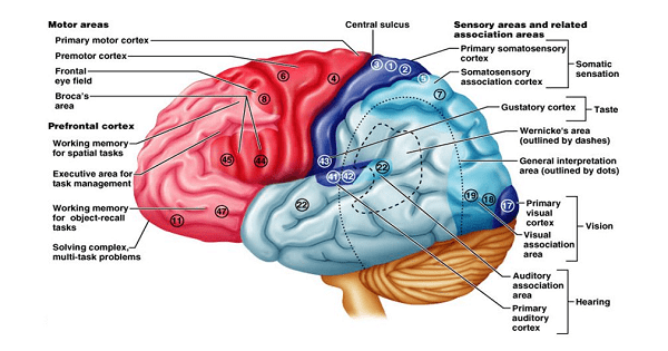
FUNCTIONAL AREAS OF CEREBRAL CORTEX
Cells present in cerebral cortex are all, except:
| A |
Bipolar cells |
|
| B |
Purkinje cells |
|
| C |
Golgi cells |
|
| D |
Granule cells |
Cells present in cerebral cortex are all, except:
| A |
Bipolar cells |
|
| B |
Purkinje cells |
|
| C |
Golgi cells |
|
| D |
Granule cells |
Bipolar cells
What is the most important source of noradrenergic innervation to the cerebral cortex?
| A |
Basal nucleus of Meynert |
|
| B |
Caudate nucleus |
|
| C |
Locus coeruleus |
|
| D |
Raphe nucleus |
What is the most important source of noradrenergic innervation to the cerebral cortex?
| A |
Basal nucleus of Meynert |
|
| B |
Caudate nucleus |
|
| C |
Locus coeruleus |
|
| D |
Raphe nucleus |
The opercular portion of the cerebral cortex that contains Broca’s area is which of the following?
| A |
Superior frontal gyrus |
|
| B |
Inferior frontal gyrus |
|
| C |
Cingulate sulcus |
|
| D |
Insula |
The opercular portion of the cerebral cortex that contains Broca’s area is which of the following?
| A |
Superior frontal gyrus |
|
| B |
Inferior frontal gyrus |
|
| C |
Cingulate sulcus |
|
| D |
Insula |
Motor speech area of Broca (Brodmann’s area 44, 45) occupies the opercular and triangular portions of the inferior frontal gyrus of the dominant hemisphere. Broca’s area is formed of two minor gyri, called pars opercularis and pars triangularis, that lie in the posterior end of the inferior frontal gyrus. This area subserves expressive language function.
Which of the area in cerebral cortex represents the frontal eye field?
| A |
Area 8 |
|
| B |
Area 6 |
|
| C |
Area 4 |
|
| D |
Area 45 |
Which of the area in cerebral cortex represents the frontal eye field?
| A |
Area 8 |
|
| B |
Area 6 |
|
| C |
Area 4 |
|
| D |
Area 45 |
Which of the following manifestation occur secondary to ablation of the ‘somatosensory area I’ of the cerebral cortex?
| A |
Total loss of pain sensation |
|
| B |
Total loss of touch sensation |
|
| C |
Loss of tactile localization but not of two point discrimination |
|
| D |
Loss of tactile localization and two point Discrimination |
Which of the following manifestation occur secondary to ablation of the ‘somatosensory area I’ of the cerebral cortex?
| A |
Total loss of pain sensation |
|
| B |
Total loss of touch sensation |
|
| C |
Loss of tactile localization but not of two point discrimination |
|
| D |
Loss of tactile localization and two point Discrimination |
Widespread bilateral excision of somatosensory area I result in person being unable to localize discretely the different sensations in the different parts of the body.
Somatosensory area 1 is located in the posterior bank of the central sulcus and on the crown of postcentral sulcus. This area corresponds to Brodmann’s area 3a, 3b, 1 and 2. Neurons in area 1 and 3b are responsive to cutaneous inputs both rapidly and slowly adapting. Area 2 and 3a respond to deep stimuli, with area 3a responsive to muscle afferents and area 2 to joints.
Area 3b is critical for the performance of tactile discrimination based on shape or texture of a stimulus. Inactivation of this area leads to lack of appreciation of the quality or existence of tactile stimuli. Lesion of area 1 disrupt performance based on texture but leaves intact performance based on stimulus size, whereas lesion in area 2 produces opposite effect.
Ref: Guyton and Hall – Textbook of Medical Physiology, 10th Edition, Page 546; Ganong’s Review of Medical Physiology, 22nd Edition, Pages 139-40; Fundamental Neuroscience, Volume 1 By Larry R. Squire, 2nd Edition, Page 689
Ablation of the ‘somatosensory area I’ of the cerebral cortex leads to :
| A |
Total loss of pain sensation |
|
| B |
Total loss of touch sensation |
|
| C |
Loss of tactile localization but not of two point discrimination |
|
| D |
Loss of tactile localization and two point discrimination |
Ablation of the ‘somatosensory area I’ of the cerebral cortex leads to :
| A |
Total loss of pain sensation |
|
| B |
Total loss of touch sensation |
|
| C |
Loss of tactile localization but not of two point discrimination |
|
| D |
Loss of tactile localization and two point discrimination |
D i.e. Loss of tactile localization & two point discrimination
Functions & Lesions
Somatosensory area I (SI)
Primary somatosensory area (SI) is Brodmann’s area 1, 2, 3 situated in postcentral gyrus. Ablation of SI would lead to cortical pattern of sensory loss. Lesion in this area produce contralateral impairment of:
– TouchQ – Sterognosis
– Pressure – Barognosis
– Proprioception – Cutaneographia
– Two point discrimationQ
|
Spinal cord lesion |
Brainstem lesion |
Thalamic lesion |
Cortical lesion |
|
Five patterns are seen depending |
Loss of pain & temperature |
Hemisensory loss of |
Patients is able to recognize all |
|
upon whether there is: |
on ipsitateral face and |
all modalitiesQ |
sensation but localizes them |
|
– Complete transaction |
opposite side of bodyQ. |
|
poorly |
|
– Hemisection |
(Lateral medullary |
|
– Loss of two point |
|
– Central cord section |
syndrome)Q |
|
discriminationQ |
|
– Posterior column section |
[Mn: TOP = Temp & Pain |
|
– AsteregnosisQ (loss of touch |
|
– Anterior column section |
Opposite side] |
|
sensation) |
|
|
|
|
– Sensory inattention |
Spongiform degeneration of cerebral cortex occurs in –
| A |
Creutzfeldt-Jakob disease |
|
| B |
Subacute sclerosing panencephalitis |
|
| C |
Fatal familial insomnia |
|
| D |
Cerebral toxoplasmosis |
Spongiform degeneration of cerebral cortex occurs in –
| A |
Creutzfeldt-Jakob disease |
|
| B |
Subacute sclerosing panencephalitis |
|
| C |
Fatal familial insomnia |
|
| D |
Cerebral toxoplasmosis |
Ans. is ‘a’ i.e., Creutzfeldt-Jakob disease
o On microscopic examination, in Creutzfeldt-Jakob disease the pathognomonic finding is a spongiform transformation of the cerebral cortex and, often, deep gray matter structures (caudate, putamen); this consists of a multifocal process that results in the uneven formation of small, apparently empty, microscopic vacuoles of varying sizes within the neuropil and sometimes in the perikaryon of neurons. In advanced cases, there is severe neuronal loss, reactive gliosis, and sometimes expansion of the vacuolated areas into cystlike spaces (“status spongiosus”).
o Kuru plaques are extracellular deposits of aggregated abnormal protein; they are Congo red-positive as well as PAS-positive and occur in the cerebellum in cases of Gerstmann-Striiussler-Scheinker syndrome; they are present in abundance in the cerebral cortex in cases of variant CJD.
o In all forms of prion disease, immunohistochemical staining demonstrates the presence of proteinase-K-resistant PrPsc in tissue.
o Fatal familial insomnia does not show spongiform pathology. Instead, the most striking alteration is neuronal loss and reactive gliosis in the anterior ventral and dorsomedial nuclei of the thalamus; neuronal loss is also prominent in the inferior olivary nuclei.
Following sensory impairments occur in extensive damage to primary somatosensory area of cerebral cortex, EXCEPT:
| A |
Pressure |
|
| B |
Sensory localization |
|
| C |
Exact weight determination |
|
| D |
Pain |
Following sensory impairments occur in extensive damage to primary somatosensory area of cerebral cortex, EXCEPT:
| A |
Pressure |
|
| B |
Sensory localization |
|
| C |
Exact weight determination |
|
| D |
Pain |
Answer is D (Pain):
Lesion of the primary somatosensory area does not lead to loss in pain sensation. Involvement of the secondary somatosensory area is associated with alteration in pain sensation.
Primary Somatosensory area is (Area 3, 1, 2)
Lesions in this area produce contralateral impairment of: –
Touch Sterognosis
Pressure Barognosis
Proprioception Cutaneographia Two point discrimination
Which of the following has small representation in somatosensory area of cerebral cortex ‑
| A |
Lips |
|
| B |
Thumb/fingers |
|
| C |
Tongue |
|
| D |
Trunk |
Which of the following has small representation in somatosensory area of cerebral cortex ‑
| A |
Lips |
|
| B |
Thumb/fingers |
|
| C |
Tongue |
|
| D |
Trunk |
Ans. is ‘d’ i.e., Trunk
A distinct topographic represention of the body can be demonstrated in somatosensory area-I.
- Each side of the cortex receives sensory information from the opposite side of the body only (contralateral representation).
- The body is represented upside down (vertical) in the postcentral gyms, i.e. the face is represented at the foot of the gyrus whereas the legs and feet are represented at the top extending on the medial surface.
- Some parts of body like face (especially lips, tongue) and fingers have a proportionately large representation than the other areas like the trunk. The cortical representation of the part of the body is proportionate to its innervation density (number of sensory receptors) rather than its size.

