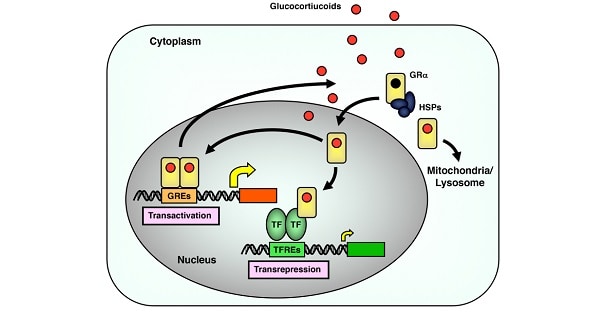
GLUCOCORTICOIDS – FUNCTIONS & REGULATION
| A | Increase in sodium and water excretion | |
| B | Increase in renal perfusion | |
| C | Decrease in cortisol levels | |
| D | Hyperkalemia |
| A | Increase in sodium and water excretion | |
| B | Increase in renal perfusion | |
| C | Decrease in cortisol levels | |
| D | Hyperkalemia |
The biochemical changes associated with shock result from tissue hypoperfusion, endocrine response to stress, and specific organ system failure. During shock, the sympathetic nervous system and adrenal medulla are stimulated to release catecholamines. Renin, angiotensin, antidiuretic hormone, adrenocorticotropin, and cortisol levels increase. Resultant changes include sodium and water retention and an increase in potassium excretion, protein catabolism, and gluconeogenesis. Potassium levels rise as a result of increased tissue release, anaerobic metabolism, and decreased renal perfusion. If renal function is maintained, potassium excretion is high and normal plasma potassium levels are restored.
Glucocorticoids causes all, except :
| A |
Muscle wasting |
|
| B |
Osteoporosis |
|
| C |
Diabetes mellitus |
|
| D |
Reduced appetite |
Glucocorticoids causes all, except :
| A |
Muscle wasting |
|
| B |
Osteoporosis |
|
| C |
Diabetes mellitus |
|
| D |
Reduced appetite |

Cortisol increases all of the following components, EXCEPT:
| A |
Platelets |
|
| B |
RBC’s |
|
| C |
Eosinophils |
|
| D |
Monocytes |
Cortisol increases all of the following components, EXCEPT:
| A |
Platelets |
|
| B |
RBC’s |
|
| C |
Eosinophils |
|
| D |
Monocytes |
The glucocorticoids are cortisol and corticosterone. The glucocorticoids decrease the number of circulating eosinophils by increasing their sequestration in the spleen and lung. Glucocorticoids also lower the number of basophils in the circulation and increase the number of neutrophils, platelets and RBC.
The typical effect of cortisol on the white and red blood cell counts in human (cells/microlitre):
|
Cell |
Normal |
Cortisol-Treated |
|
Total WBC |
9000 |
10000 |
|
PMNs |
5760 |
8330 |
|
Lymphocytes |
2370 |
1080 |
|
Eosinophils |
270 |
20 |
|
Basophils |
60 |
30 |
|
Monocytes |
450 |
540 |
Ref: Barrett K.E., Barman S.M., Boitano S., Brooks H.L. (2012). Chapter 20. The Adrenal Medulla & Adrenal Cortex. In K.E. Barrett, S.M. Barman, S. Boitano, H.L. Brooks (Eds), Ganong’s Review of Medical Physiology, 24e.
Which of the following areas of the adrenal gland would you expect to increase in activity in a patient subjected to salt restriction?
| A |
Adrenal medulla |
|
| B |
Zona fasciculata of the adrenal cortex |
|
| C |
Zona glomerulosa of the adrenal cortex |
|
| D |
Zona reticularis of the adrenal cortex |
This question requires you to equate salt restriction with an increased synthesis of aldosterone (aldosterone promotes sodium reabsorption) and then to remember that aldosterone is produced in the zona glomerulosa of the adrenal cortex. The zona glomerulosa is the outermost layer of the adrenal cortex.
The adrenal medulla secretes catecholamines.
The zona fasciculata is the middle layer of the adrenal cortex. It primarily secretes glucocorticoids.
The zona reticularis is the innermost layer of the adrenal cortex. It primarily secretes androgens such as dehydroepiandrosterone (DHEA).
Ref: Barrett K.E., Barman S.M., Boitano S., Brooks H.L. (2012). Chapter 20. The Adrenal Medulla & Adrenal Cortex. In K.E. Barrett, S.M. Barman, S. Boitano, H.L. Brooks (Eds), Ganong’s Review of Medical Physiology, 24e.
During stress increased stimulation of cortisol release cause :
| A |
Neoglucogenesis |
|
| B |
Glycolysis |
|
| C |
Protein breakdown |
|
| D |
A & C |
During stress increased stimulation of cortisol release cause :
| A |
Neoglucogenesis |
|
| B |
Glycolysis |
|
| C |
Protein breakdown |
|
| D |
A & C |
A i e. Neoglucogenesis C i.e. Protein breakdown
It is a catabolic hormone. Their function appears to be oriented to maintain blood glucose levels during starvation – so that brain continues to get its nutrients.
* In stress vasopressinQ also increases Glucocorticoids
* Glucocorticoids inhibit intestinal absorption & enhance renal excretion of Ca++
* In adrenal insufficiency, the blood glucose is normal as long as food intake is maintained, but fasting precipitates hypoglycemia & collapse. This is because glucocorticoids are necessary for glucagons to exert its gluconeogenic action during fasting.
Anti inflammatory action of cortisol is due to?
| A |
Effect on lymphocytes |
|
| B |
Effect on cytokines |
|
| C |
Effect on enzymes |
|
| D |
All of the above |
Anti inflammatory action of cortisol is due to?
| A |
Effect on lymphocytes |
|
| B |
Effect on cytokines |
|
| C |
Effect on enzymes |
|
| D |
All of the above |
Ans. is ‘d’ i.e., All of the above
Glucocorticoids act in inflammation by ‑
| A |
↓ Lipocortin |
|
| B |
↑ IL2 |
|
| C |
↑ Lipocortin |
|
| D |
↑ CRP |
Glucocorticoids act in inflammation by ‑
| A |
↓ Lipocortin |
|
| B |
↑ IL2 |
|
| C |
↑ Lipocortin |
|
| D |
↑ CRP |
Ans. is ‘c’ i.e., ↑ Lipocortin
o Corticosteroids induce the production of lipocortins that are responsible for the inhibition of phosholipase A2, an enzyme which is involved in the production of inflammatory mediators (PGs, LTs, PAF).
Glucocorticoids do not cause?
| A |
Osteoporosis |
|
| B |
Hypoglycemia |
|
| C |
Peptic ulceration |
|
| D |
Cataracts |
Glucocorticoids do not cause?
| A |
Osteoporosis |
|
| B |
Hypoglycemia |
|
| C |
Peptic ulceration |
|
| D |
Cataracts |
Ans. is ‘b’ i.e., Hypoglycemia
o Glucocorticoids cause hyperglycemia.
o All other are side effects of glucocorticoids
o Side effects of glucocorticoids are cuhing’s habits, fragile skin, purple striae, muscular weakness, hyperglycemia, delayed wound healing hypokalemia, Na+ and water retention, hypertension, peptic ulcer, cataract, glaucoma, osteoporosis, mild euphoria, growth retardation and AVN of femoral head. Among cutaneous side effects are folliculitis (most common), atrophic changes, hypertrichosis, and purple stria are common.
Long term administration of glucocorticoids can cause all of the following except‑
| A |
Proximal myopathy |
|
| B |
Hyperkalemia |
|
| C |
Hypertension |
|
| D |
Cataract |
Long term administration of glucocorticoids can cause all of the following except‑
| A |
Proximal myopathy |
|
| B |
Hyperkalemia |
|
| C |
Hypertension |
|
| D |
Cataract |
Ans. is ‘b’ i.e., Hyperkalemia
o Glucocorticoids have some mineralocorticoid activity, thus they cause hypokalemia (not hyperkalemia).
The first reactionary change to occur after vessel injury and haemorrhage is:
September 2011
| A |
Vasoconstriction |
|
| B |
Bradycardia |
|
| C |
Raised cortisol |
|
| D |
Raised adrenaline |
The first reactionary change to occur after vessel injury and haemorrhage is:
September 2011
| A |
Vasoconstriction |
|
| B |
Bradycardia |
|
| C |
Raised cortisol |
|
| D |
Raised adrenaline |
Ans. A: Vasoconstriction
When a small blood vessel is transected or damaged, the injury initiates a series of events that lead to the formation of a clot. This seals off the damaged region and prevents further blood loss.
The initial event is constriction of the vessel
Physiological response to hemorrhage
- It is a three-part process. The principal features are:
– Rapidly responding neural and humoral mechanisms direct available blood flow toward vital organs.
– More slowly evolving salt and water retention by the kidneys replaces the lost plasma.
– Erythropoiesis gradually replaces the lost red blood cells.
Acute response to hemorrhage
- The acute response includes a primary decrease in cardiac output, a secondary decrease in arterial pressure and compensatory increases in heart rate and vascular resistance.
Autonomic involvement
- Increased heart rate and peripheral vasoconstriction signal the autonomic nervous system’s participation in the acute response to hemorrhage.
- Autonomic dysfunction decreases the body’s tolerance to blood loss.
Selective vasoconstriction
- Sympathetic vasoconstriction during hemorrhage is selective.
- Many organs show intense vasoconstriction, while the cerebral and coronary circulations show little response to increased sympathetic outflow.
- The benefit of selectivity is that available blood flow, as meager as it may be, is preferentially directed to the brain and heart — the vital organs.
Support from Renin-Angiotensin system
- The renin-angiotensin system supports arterial pressure in hemorrhage by constricting nonvital organs.
- This response is slower than the response of the sympathetic nervous system, but it is still an important part of the acute circulatory response to hemorrhage.
Salt and water retention
- After hemorrhage, with no intervention, salt and water retention over several days will increase blood volume to normal or above.
- Erythrocytes are replaced much more slowly.
Regarding therapeutic uses of steroids, which of the following is not properly matched:
| A |
Dexamethasone – Cerebral edema |
|
| B |
Betamethasone – Cerebral edema |
|
| C |
Prednisolone – Rheumatoid arthritis |
|
| D |
Cortisol – Cushings syndrome |
Regarding therapeutic uses of steroids, which of the following is not properly matched:
| A |
Dexamethasone – Cerebral edema |
|
| B |
Betamethasone – Cerebral edema |
|
| C |
Prednisolone – Rheumatoid arthritis |
|
| D |
Cortisol – Cushings syndrome |
Ans. D: Cortisol – Cushings syndrome
Amongst endogenous causes, pituitary dependent cortisol excess (by convention, called Cushing’s disease) accounts for approximately 80% of the cases of Cushings syndrome