
HEMOGLOBINOPATHIES
| A | Inherited as AR | |
| B | Inherited as AD | |
| C | Inherited as XLR | |
| D | Inherited as XLD |
| A | Inherited as AR | |
| B | Inherited as AD | |
| C | Inherited as XLR | |
| D | Inherited as XLD |
Sickle cell trait patient do not have manifestations as that of Sickle cell disease, because:
| A |
50% HbS is required for occurrence of sickling |
|
| B |
HbA prevents sickling |
|
| C |
50% sickles |
|
| D |
HbA prevent polymerization of Hbs |
Sickle cell trait patient do not have manifestations as that of Sickle cell disease, because:
| A |
50% HbS is required for occurrence of sickling |
|
| B |
HbA prevents sickling |
|
| C |
50% sickles |
|
| D |
HbA prevent polymerization of Hbs |
What is the cause of sickling of RBC in sickle cell disease?
| A |
Decreased Solubility |
|
| B |
Decreased Stability |
|
| C |
Altered Function |
|
| D |
Altered 02 binding capacity |
What is the cause of sickling of RBC in sickle cell disease?
| A |
Decreased Solubility |
|
| B |
Decreased Stability |
|
| C |
Altered Function |
|
| D |
Altered 02 binding capacity |
In sickle cell disease glutamic acid is replaced by valine.
The charge and location of this substitution cause HbS to be converted from a soluble state into a polymer when it undergoes structural changes that accompany release of oxygen. Hypoxia, acidosis, and hypertonicity facilitate polymer formation.
The polymerization of hemoglobin cause the red cell to transform from a deformable, biconcave disk into a rigid, sickle shaped cell.
Ref: Rudolph’s Fundamentals of Pediatrics By Abraham M. Rudolph, Pages 528-30

True about Sickle cell disease are all, Except:
| A |
Single nucleotide change results in change of Glutamine to Valine |
|
| B |
Deoxygenated Hb 1/t exposure of sticky end d/ t replacement of nonpolar residue by polar residue |
|
| C |
HbS confers resistance against malaria in heterozygotes |
|
| D |
RFLP results from a single base change |
True about Sickle cell disease are all, Except:
| A |
Single nucleotide change results in change of Glutamine to Valine |
|
| B |
Deoxygenated Hb 1/t exposure of sticky end d/ t replacement of nonpolar residue by polar residue |
|
| C |
HbS confers resistance against malaria in heterozygotes |
|
| D |
RFLP results from a single base change |
B i.e. Deoxygenated Hb Vt exposure of sticky end d/t replacement of nonpolar residue by polar residue
All of the following are true about Sickle cell disease, Except:
| A |
Single nucleotide change results in change of Glutamine to Valine |
|
| B |
RFLP results from a single base change |
|
| C |
‘Sticky patch’ is generated as a result of replacement of a non polar residue with a polar residue |
|
| D |
HbS confers resistance against malaria in heterozygotes |
All of the following are true about Sickle cell disease, Except:
| A |
Single nucleotide change results in change of Glutamine to Valine |
|
| B |
RFLP results from a single base change |
|
| C |
‘Sticky patch’ is generated as a result of replacement of a non polar residue with a polar residue |
|
| D |
HbS confers resistance against malaria in heterozygotes |
C i.e. ‘Sticky patch’ is generated as a result of replacement of a non polar residue with a polar residue
In sickle cell anemia- Glutamic acid is replaced by valine at sixth position in /3 – globin chainQ.
Sticky patch is generated as a result of replacement of polar residue (negatively charged glutamate amino acid) with a nonpolar hydrophobic residue (valine)Q. This change makes HbS (sickle cell hemoglobin) less soluble, when deoxygenated and cause sicklingQ.
HbS confers resistance against malaria in heterozygotesQ.
Sickle cell trait patient do not have manifestations as that of Sickle cell disease, because‑
| A |
50% HbS is required for occurrence of sickling |
|
| B |
HbA prevents sickling |
|
| C |
50% sickles |
|
| D |
HbA prevents polymerazitation of Hbs |
Sickle cell trait patient do not have manifestations as that of Sickle cell disease, because‑
| A |
50% HbS is required for occurrence of sickling |
|
| B |
HbA prevents sickling |
|
| C |
50% sickles |
|
| D |
HbA prevents polymerazitation of Hbs |
Ans. is ‘a’ i.e., 50% Hbs is required for occurence of sickling
- Sickle cell disease occurs in following forms
1. Heterozygous state, i.e., sickle cell trait.
2. Homozygous state, i.e., sickle cell anemia.
o The most important factor for sickling is the amount of HbS in the cell.
o In sickle cell trait (heterozygous state), only about 40% of the hemoglobin is HbS, this is insufficient to produce sickling manifestations.
o In homozygous state, almost all the hemoglobin in the red cell is HbS, therefore they undergo sickling.
September 2007
| A |
Glutamtic acid, at position No.5 of beta-globin chain of haemoglobin is replaced by valine |
|
| B |
Glutamtic acid, at position No.6 of beta-globin chain of haemoglobin is replaced by valine |
|
| C |
Valine at position No.6 of beta-globin chain of haemoglobin is replaced by glutamtic acid |
|
| D |
Valine at position No.5 of beta-globin chain of haemoglobin is replaced by glutamtic acid |
In sickle cell disease:
September 2007
| A |
Glutamtic acid, at position No.5 of beta-globin chain of haemoglobin is replaced by valine |
|
| B |
Glutamtic acid, at position No.6 of beta-globin chain of haemoglobin is replaced by valine |
|
| C |
Valine at position No.6 of beta-globin chain of haemoglobin is replaced by glutamtic acid |
|
| D |
Valine at position No.5 of beta-globin chain of haemoglobin is replaced by glutamtic acid |
Ans. B: Glutamtic acid, at position No.6 of beta-globin chain of haemoglobin is replaced by valine
This happens due to change of a nucleotide, adenine to thymine (GAGgGTG) of codon 6 of beta-globin gene. This substitution of amino acid changes the net charge of haemoglobin, oxygen affinity and three-dimensional structure thus rendering it as unstable haemoglobin.
Sickle haemoglobin gets polymerized at low oxygen tension and deforms the red blood cell from discoid shape to sickle like (crescent) form
Person having heterozygous sickle cell trait is protected from infection of:
| A |
P. falciparum |
|
| B |
P. vivax |
|
| C |
Pneumococcus |
|
| D |
Salmonella |
Person having heterozygous sickle cell trait is protected from infection of:
| A |
P. falciparum |
|
| B |
P. vivax |
|
| C |
Pneumococcus |
|
| D |
Salmonella |
Ans. a. P. falciparum
- Person having heterozygous sickle cell trait is protected from infection of P. falciparum.
- “People who are heterozygous for the sickle cell trait (HbS) become infected with P. falciparum, but they are less likely to die from infection°. The HbS trait causes the parasites to grow poorly or die because of the low oxygen concentrations°.” – Robbins 8/e p387
Host Resistance to Plasmodium
- Two general mechanisms of host resistance to Plasmodium:
- Inherited alterations in red cells make people resistant to Plasmodium°.
- Repeated or prolonged exposure to Plasmodium species stimulates an immune response that reduces the severity
- People who are heterozygous for the sickle cell trait (HbS) become infected with P. falciparum, but the yare less likely to die from infection°.
- The HbS trait causes the parasites to grow poorly or die because of the low oxygen concentrations°.
- The geographic distribution of the HbS trait is similar to that of P. falciparum°, suggesting evolutionary selection of the HbS trait in people by the parasite
- HbC, another common hemoglobin mutation, also protects against severe malaria by reducing parasite proliferation°.
Host Resistance to Plasmodium.
- People can also be resistant to malaria due to the absence of proteins to which the parasites bind°.
- P. vivax enters red cells by binding to the Duffy blood group antigen°.
- Many Africans, including most Gambians, are not susceptible to infection by P. vivax because they do not have the Duffy antigen°.
- Antibodies and T lymphocytes specific for Plasmodium reduce disease manifestations. Cytotoxic lymphocytes may also be important in resistance to P. falciparum
| A |
Decreased |
|
| B |
Increased |
|
| C |
Dependent on HbS concentration |
|
| D |
Unchanged. |
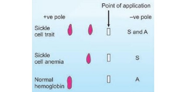
After a point mutation, glutamic acid is replaced by valine, which leads to formation of sickle cell hemoglobin. The mobility of HbS as compared with normal hemoglobin on gel electrophoresis will be:
| A |
Decreased |
|
| B |
Increased |
|
| C |
Dependent on HbS concentration |
|
| D |
Unchanged. |
Ans: A. Decreased
(Ref: Harper 30/e p460, 28/e p49, 357. 3197-398; Lippincott 5/e p|75-177; Lehinger 5/e pl(t8-l69t)
HbS mobility on gel electrophoresis:
- Decreased mobility – Compared to normal hemoglobin.
Electrophoresis of hemoglobin:
- Obtained from lysed red blood cells.
- Used in sickle cell trait & disease diagnosis.
Sequence of Movement: HbA2 < HbC < HhS < HbF < HbA

