
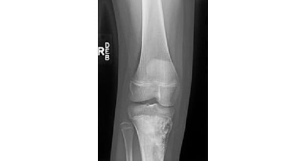
OSTEOSARCOMA (OSTEOGENIC SARCOMA)
Which of the following tumor is least radiosensitive hence it is ineffective for radiotherapy?
| A |
Ewing’s sarcoma |
|
| B |
Osteosarcoma |
|
| C |
Wilms tumor |
|
| D |
Neuroblastoma |
Which of the following tumor is least radiosensitive hence it is ineffective for radiotherapy?
| A |
Ewing’s sarcoma |
|
| B |
Osteosarcoma |
|
| C |
Wilms tumor |
|
| D |
Neuroblastoma |
A 45 year old man presents to a physician with back pain and facial pain. Physical examination demonstrates coarse facial features and kyphosis. Laboratory examination is remarkable for elevated alkaline phosphatase. .X-ray studies demonstrate skull thickening with narrowing of foramina, and bowing of the femur and tibia. Bone biopsy reveals a mosaic pattern of bone spicules with prominent osteoid seams. Which of the following neoplasms occurs at an increased frequency in patients with this disorder?
| A |
Astrocytoma |
|
| B |
Hodgkin’s lymphoma |
|
| C |
Meningioma |
|
| D |
Osteosarcoma |
A 45 year old man presents to a physician with back pain and facial pain. Physical examination demonstrates coarse facial features and kyphosis. Laboratory examination is remarkable for elevated alkaline phosphatase. .X-ray studies demonstrate skull thickening with narrowing of foramina, and bowing of the femur and tibia. Bone biopsy reveals a mosaic pattern of bone spicules with prominent osteoid seams. Which of the following neoplasms occurs at an increased frequency in patients with this disorder?
| A |
Astrocytoma |
|
| B |
Hodgkin’s lymphoma |
|
| C |
Meningioma |
|
| D |
Osteosarcoma |
15 year old boy presented with a mass in the distal femur. X-ray from the lesion showed features of Codman’s triangle and Sunray appearance. The diagnosis is:
| A |
Osteosarcoma |
|
| B |
Ewing’s sarcoma |
|
| C |
Osteoclastoma |
|
| D |
Chondroblastoma |
15 year old boy presented with a mass in the distal femur. X-ray from the lesion showed features of Codman’s triangle and Sunray appearance. The diagnosis is:
| A |
Osteosarcoma |
|
| B |
Ewing’s sarcoma |
|
| C |
Osteoclastoma |
|
| D |
Chondroblastoma |
Codman’s triangle and Sunray appearance are characteristic radiological findings in osteosarcoma.
When the tumour elevates the periosteum of bone, a gap occurs between periosteum and bone, where the gap is elevated, bone is formed, creating a triangular shape on radiographs.
Ref: Apley’s system of orthopaedics and fractures;8th/185; Essential orthopedics by Maheshwari 3rd/219, 222.

Which of the following investigations is not required in Osteosarcoma?
| A |
MRI |
|
| B |
Bone marrow biopsy |
|
| C |
CT chest |
|
| D |
Bone scan |
Which of the following investigations is not required in Osteosarcoma?
| A |
MRI |
|
| B |
Bone marrow biopsy |
|
| C |
CT chest |
|
| D |
Bone scan |
A biopsy from the lesion is essential for grading of the tumour but a biopsy from bone marrow is not indicated.
Bone scan, CT chest and MR1 femur are important staging investigations for osteosarcoma.
Ref: Apley’s system of orthopaedics and fractures 8th/185; Pediatric and Adolescent Osteosarcoma By Norman Jaffe, Øyvind S. Bruland, 2009, Page 209; Final FRCR 2B Long Cases: A Survival Guide By Jessie Aw, John Curtis, 2010, Page 165.
Which of the following is a ‘Pulsating tumor’ of the bone?
| A |
Osteosarcoma |
|
| B |
Chondrosarcoma |
|
| C |
Ewing’s sarcoma |
|
| D |
Eosinophilic Granuloma |
Which of the following is a ‘Pulsating tumor’ of the bone?
| A |
Osteosarcoma |
|
| B |
Chondrosarcoma |
|
| C |
Ewing’s sarcoma |
|
| D |
Eosinophilic Granuloma |
A child with a history of pain over lower extremities since few moths shows radiological evidence of osteosarcoma. Most common site of osteosarcoma is:
| A |
Upper end of femur |
|
| B |
Lower end of femur |
|
| C |
Lower end of fibula |
|
| D |
Lower end of tibia |
A child with a history of pain over lower extremities since few moths shows radiological evidence of osteosarcoma. Most common site of osteosarcoma is:
| A |
Upper end of femur |
|
| B |
Lower end of femur |
|
| C |
Lower end of fibula |
|
| D |
Lower end of tibia |
Second most common malignancy in a patient with retinoblastoma is:
| A |
Nephroblastoma |
|
| B |
Osteosarcoma |
|
| C |
Neuroblastoma |
|
| D |
Craniopharyngioma |
Second most common malignancy in a patient with retinoblastoma is:
| A |
Nephroblastoma |
|
| B |
Osteosarcoma |
|
| C |
Neuroblastoma |
|
| D |
Craniopharyngioma |
Retinoblastoma:
- It is the most common primary intraocular malignancy of childhood.
- The mutation occurs in the retinoblastoma gene (Rb) at chromosome 13q14. This is a tumor suppressor gene.
- One mutated copy may be inherited in an autosomal dominant fashion (germline mutation). If a second mutation spontaneously occurs in any cell, tumorigenesis is likely.
- Individuals with a germinal mutation are at risk for the development of tumors other than retinoblastoma (pineal tumors, osteosarcoma, and other soft tissue sarcomas).
A patient with which of the follwing disease is predisposed to develop osteosarcoma –
| A |
Osteomalacia |
|
| B |
Osteomyelitis |
|
| C |
Paget’s disease of bone |
|
| D |
b and c |
A patient with which of the follwing disease is predisposed to develop osteosarcoma –
| A |
Osteomalacia |
|
| B |
Osteomyelitis |
|
| C |
Paget’s disease of bone |
|
| D |
b and c |
Ans. is ‘b’ i.e., Osteomyelitis & c i.e., Paget’s disease of bone
Osteosarcoma (osteogenic sarcoma – OGS)
o Osteosarcoma is primary malignant bone tumor of bone, derived from primitive bone forming mesenchyma and characterized by osteoid formation.
- It is of two types
- Primary
- Secondary
o Paget disease
o Radiation
o Fibrous dysplasia
o Enchondromatosis
o Multiple osteochondroma
o Bone infarction
o Chronic osteomyelitis
Which of the following is not true in relation to osteosarcoma?
| A |
Paget’s disease & prior irradiation are predisposing factors |
|
| B |
Rb gene mutation is associated with hereditary variant |
|
| C |
c-myc gene implicated in the genesis |
|
| D |
Codman’s triangle is the characteristic x-ray finding |
Which of the following is not true in relation to osteosarcoma?
| A |
Paget’s disease & prior irradiation are predisposing factors |
|
| B |
Rb gene mutation is associated with hereditary variant |
|
| C |
c-myc gene implicated in the genesis |
|
| D |
Codman’s triangle is the characteristic x-ray finding |
Ans. is c i.e., c-myc gene implicated in the genesis
o Genetic mutations are fundamental to the development of osteosarcoma. Patients with herediatary retinoblastomas have upto 1000 times greater risk of subsequently developing osteosarcoma, attributed to germline mutation in RB gene. Abnormalities in gene that regulate cycling such as p53, CDK4, p16, INK4A, CYCLINDI and MDM2 have also been implicated in the genesis of nonherediatary osteosarcoma.
Radiological features of osteosarcoma
o X-ray of a patient with osteosarcoma shows following features : –
1)An area of irregular destruction in the metaphysis, sometimes overshadowed by the new bone formation. The
cortex overlying the lesion is eroded. There is new bone formation in the matrix of the tumour.
2)Periosteal reaction : – As the tumor lifts the periosteum, it incites an intense periosteal reaction. The periosteal
reaction in an osteosarcoma is irregular, unlike in osteomyelitis where it is smooth and in layers.
3)Codman’s triangle: – A triangular area of subperiosteal new bone is seen at the tumour host cortex junction at the
ends of the tumor.
4)Sun-ray appearance:- As the periosteum is unable to contain the tumour, the tumour grows into the overlying soft tissues. New bone is laid down along the blood vessels within the tumour growing centrifugally, giving rise to a `sun-ray appearance’ on the X-ray.
Radiological feature of osteosarcoma is:
| A |
New bone formation |
|
| B |
Sunray appearance |
|
| C |
Cotton wool app. |
|
| D |
Osteoid formation |
Radiological feature of osteosarcoma is:
| A |
New bone formation |
|
| B |
Sunray appearance |
|
| C |
Cotton wool app. |
|
| D |
Osteoid formation |
B i.e. Sunray appearance
The X Ray findings of osteosarcomas are characterized by:
Variable mixture of radiopacities (due to osteogenesis) & radiolucencies (due to destructive change & replacement by radiolucent osteoid)
– Sun Burst/ Sun ray appearanceQ
– Codman’s triangleQ (also found in infection & Ewing’s SA so not specific)
|
Disease |
Radiological Feature |
|
Osteosarcoma |
• Variable mixture of radiopacities & radiolucency (hallmark)(2 |
|
|
• Sun burst/sun ray appearanceQ (IInd) |
|
|
• Codman’s triangleQ (non specific) |
|
Osteoclastoma |
Soap Bubble appearanceQ |
|
Ewing’s Sarcoma |
Onion peel appearanceQ |
|
Paget’s disease |
• In Osteolytic Hot phase |
|
|
– Advancing wedge/Blade of Grass/Candle flame appearance |
|
|
(Mn = ABC) |
|
|
• In Mixed phase |
|
|
– Cotton Ball SkullQ |
|
|
– Picture Frame VertebraeQ |
|
|
• In Cool Phase |
|
|
– Bowing |
|
|
– Bone density increased |
Osteosarcoma commonly affects:
September 2005
| A |
Metaphysis |
|
| B |
Diaphysis |
|
| C |
Epiphysis |
|
| D |
None of the above |
Osteosarcoma commonly affects:
September 2005
| A |
Metaphysis |
|
| B |
Diaphysis |
|
| C |
Epiphysis |
|
| D |
None of the above |
Ans. A: Metaphysis
Metaphysis-common site for osteosarcoma
- Diachysis- common site for ewing’s tumor
- Epiphysis- common site for osteoclastoma
Osteosarcoma is differentiated from myositis ossificans by radiology by:
| A |
Location |
|
| B |
Absence of osteomyelitic changes |
|
| C |
Shape of swelling |
|
| D |
Peripheral field of differentiation |
Osteosarcoma is differentiated from myositis ossificans by radiology by:
| A |
Location |
|
| B |
Absence of osteomyelitic changes |
|
| C |
Shape of swelling |
|
| D |
Peripheral field of differentiation |
Ans. Peripheral field of differentiation
Which of the following is not sensitive to radiotherapy?
| A |
Ewings sarcoma |
|
| B |
Osteosarcoma |
|
| C |
Wilms’ |
|
| D |
Neuroblastoma |
Which of the following is not sensitive to radiotherapy?
| A |
Ewings sarcoma |
|
| B |
Osteosarcoma |
|
| C |
Wilms’ |
|
| D |
Neuroblastoma |
Ans. Osteosarcoma
The given x-ray shows the presence of codman’s triangle which is is seen in cases of osteosarcoma.Calcification in osteosarcoma is due to presence of ?

| A |
Osteoid matrix |
|
| B |
Osteoblasts |
|
| C |
High Calcium levels in serum |
|
| D |
High calcitonin |
The given x-ray shows the presence of codman’s triangle which is is seen in cases of osteosarcoma.Calcification in osteosarcoma is due to presence of ?
| A |
Osteoid matrix |
|
| B |
Osteoblasts |
|
| C |
High Calcium levels in serum |
|
| D |
High calcitonin |
Matrix mineralization (calcification) of tumor matrix
- The pattern of mineralization (calcification) on radiograph may be helpful in identifying tumor matrix.
- Dense, homogenous mineralization (calcification) is typical of osteoid matrix, formed by benign and malignant bone-forming lesions.However, osteoid matrix is not always mineralized.
- Calcified rings and arcs, dense punctate calcification, and flocculent calcification (small, loosely aggregated masses) are pattern of mineralization of chondroid matrix, formed by benign and malignant cartilage forming tumors.
Identify the tumor in this patient complainig of pain immediately below the knee worse at night, intermittent and of varying intensity

| A |
Osteosarcoma |
|
| B |
Osteochondroma |
|
| C |
Chondrosarcoma |
|
| D |
Fibrosarcoma |
Identify the tumor in this patient complainig of pain immediately below the knee worse at night, intermittent and of varying intensity
| A |
Osteosarcoma |
|
| B |
Osteochondroma |
|
| C |
Chondrosarcoma |
|
| D |
Fibrosarcoma |
Ans:A.)Osteosarcoma
The condition shown in the picture above represents Osteogenic sarcoma.
Osteogenic sarcoma (osteosarcoma)
- It is the most common primary malignant tumor of bone, excluding plasma cell myeloma.
- Osteosarcoma is a bone tumor and can occur in any bone, usually in the extremities of long bones near metaphyseal growth plates. The most common sites are as follows:
- Femur ,Tibia, Humerus ,Skull and jaw, Pelvis
- Radiograph of the femur in a patient with osteosarcoma shows a typical Codman triangle ( A characteristic often seen in an X-ray is Codman’s triangle(which is basically a subperiosteal lesion formed when the periosteum is raised due to the tumor) and more diffuse, mineralized osteoid within the soft tissues adjacent to the bone.
- Symptoms include pain worse at night, intermittent and of varying intensity .
- It is treated by a combination of surgical excision and chemotherapy.
Diagnose the condition as “marked by a red arrow” in the picture below ?

| A |
Chondroblastoma. |
|
| B |
Chondrosarcoma. |
|
| C |
Osteosarcoma. |
|
| D |
Giant cell tumour. |
Diagnose the condition as “marked by a red arrow” in the picture below ?
| A |
Chondroblastoma. |
|
| B |
Chondrosarcoma. |
|
| C |
Osteosarcoma. |
|
| D |
Giant cell tumour. |
The arrow marked in the picture above represents the Codman’s triangle.
Codman triangle is the triangular area of new subperiosteal bone that is created when a lesion, often a tumour, raises the periosteum away from the bone. A Codman triangle is not actually a full triangle. Instead it is often a pseudotriangle on radiographic findings with ossification on the original bone and one additional side of the triangle which forms a two sided triangle with one open side. This two sided appearance is generated due to a tumor (or growth) that is growing at a rate which is faster than the periosteum can grow or expand, so instead of dimpling, the periosteum tears away and provides ossification on the second edge of the triangle.
The Codman triangle may be seen with aggressive lesions:
- Osteosarcoma.
- Ewing sarcoma.
- Osteomyelitis.
- Active aneurysmal bone cyst.
- Giant cell tumor.
A child is diagnosed with osteosarcoma based on sunray appearance on X-ray. This is because of:
| A |
Calcification along the periosteum |
|
| B |
Calcification along the blood vessels |
|
| C |
Periosteal reaction |
|
| D |
Soft tissue invasion |
A child is diagnosed with osteosarcoma based on sunray appearance on X-ray. This is because of:
| A |
Calcification along the periosteum |
|
| B |
Calcification along the blood vessels |
|
| C |
Periosteal reaction |
|
| D |
Soft tissue invasion |
Ans. c. Periosteal reaction

