
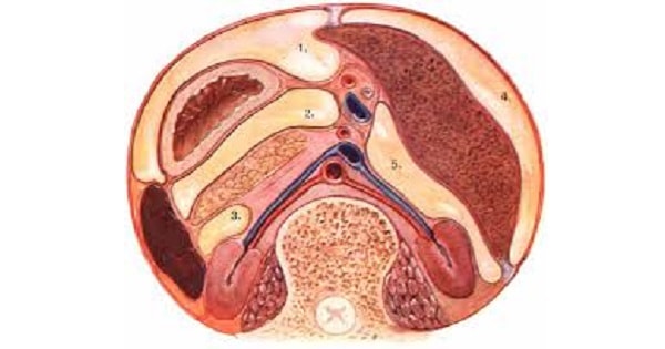
Sub Phrenic Abcess
38.20C and he has decreased breath sounds at left lung base. His abdominal wound appears to be healing well, bowel sounds are active and there are no peritoneal signs. Digital rectal examination is negative. WBC 12,500/mm3 with a shift to left. CXR show ‘plate like’ atelectasis of the left lung field. Abdominal radiograph shows a non-specific gas pattern in bowel and an air-fluid level in LUQ. Serum Amylase is 150 Somogyi units per dL. The most likely diagnosis is:
| A | Subphrenic abscess | |
| B | Subfascial wound infection | |
| C | Pancreatitis | |
| D | Pulmonary embolism |
38.20C and he has decreased breath sounds at left lung base. His abdominal wound appears to be healing well, bowel sounds are active and there are no peritoneal signs. Digital rectal examination is negative. WBC 12,500/mm3 with a shift to left. CXR show ‘plate like’ atelectasis of the left lung field. Abdominal radiograph shows a non-specific gas pattern in bowel and an air-fluid level in LUQ. Serum Amylase is 150 Somogyi units per dL. The most likely diagnosis is:
| A | Subphrenic abscess | |
| B | Subfascial wound infection | |
| C | Pancreatitis | |
| D | Pulmonary embolism |
(Subphrenic abscess)
Following Laparotomy it has been established that half of all patients develop some postoperative pulmonary collapse. This patient has pain in epigastrium, which exacerbates by deep breathing; hence, he will have some respiratory compromise adding to retained secretions.Leucocytosis, epigastric pain, local paralytic ileus in LUQ in the form of an air-fluid level there, with the reduced breath sounds at LLZ of lungs in a postoperative patient direct towards a infective subdiaphragmatic pathology, in turn affecting LLZ in form of Fleischner’s plate atelectasis, i.e., most likely to be subphrenic abscess. However, the subsegmental or plate-like atelectasis as such appears about 24 hrs post-operatively and resolves in 2–3 days. Here the subdiaphragmatic abscess would be important.
Ten days after a splenectomy for blunt abdominal trauma, a 23-year-old man complains of upper abdominal and lower chest pain exacerbated by deep breathing. He is anorectic but ambulatory and otherwise making satisfactory progress. On physical examination, his temperature is 38.2°C (108°C) rectally, and he has decreased breath sounds at the left lung base. His abdominal wound appears to be healing well, bowel sound are active and there are no peritoneal signs. Rectal examination is negative. The W.B.C. count is 12,500 mm3 with a shift to left. Chest X-rays show platelike atelectasis of the left lung field. Abdominal X-rays show a nonspecific gas pattern in the bowel and an air-fluid level in the left upper quadrant. Serum amylase is 150 Somogyi units dl (normal 60 to 80). The most likely diagnosis is:
| A |
Subphrenic abscess |
|
| B | ||
| C |
Pancreatitis |
|
| D |
Pulmonary embolism |
Ten days after a splenectomy for blunt abdominal trauma, a 23-year-old man complains of upper abdominal and lower chest pain exacerbated by deep breathing. He is anorectic but ambulatory and otherwise making satisfactory progress. On physical examination, his temperature is 38.2°C (108°C) rectally, and he has decreased breath sounds at the left lung base. His abdominal wound appears to be healing well, bowel sound are active and there are no peritoneal signs. Rectal examination is negative. The W.B.C. count is 12,500 mm3 with a shift to left. Chest X-rays show platelike atelectasis of the left lung field. Abdominal X-rays show a nonspecific gas pattern in the bowel and an air-fluid level in the left upper quadrant. Serum amylase is 150 Somogyi units dl (normal 60 to 80). The most likely diagnosis is:
| A |
Subphrenic abscess |
|
| B | ||
| C |
Pancreatitis |
|
| D |
Pulmonary embolism |
Patient is showing features of subphrenic abcess. Patient presents with local or subcostal pain and tenderness, unexplained fever and tachycardia.
Investigation: CBC shows leucocytosis.
X-ray chest shows elevated hemidiaphragm, blunting of the costophrenic angle, pleural effusion and pulmonary infiltrates or atelectasis.
CT scan of abdomen is used to confirm the diagnosis. Scanning techniques such as gallium technetium subtraction or following injection of gallium labeled leucocytes is also useful in the localization of subphrenic abscess.
Treatment includes surgical drainage and appropriate antibiotic therapy.
Ref: Crofton and Douglas’s Respiratory Diseases: 2 Volume Set, 5th Edition By Anthony Seaton, Page 1239; Common Surgical Emergencies By S. K. Kochar, Page 139; SRB’s Manual of Surgery By Bhat, 3rd Edition, Page 508
Correct about subphrenic abscess is ‑
| A |
Rarely chest symptoms |
|
| B |
Toxaemia |
|
| C |
Rarely toxaemia |
|
| D |
No sign and symptoms |
Correct about subphrenic abscess is ‑
| A |
Rarely chest symptoms |
|
| B |
Toxaemia |
|
| C |
Rarely toxaemia |
|
| D |
No sign and symptoms |
Ans. is ‘b’ i.e., Toxaemia
A middle aged patient presents with complaint of right hypochondrial pain. On plain chest x ray, elevated right hemidiaphragm is seen. All of the following are possible diagnosis except:
| A |
Acute cholecystitis |
|
| B |
Subphrenic abscess |
|
| C |
Amebic liver abscess |
|
| D |
Pyogenic liver abscess |
A middle aged patient presents with complaint of right hypochondrial pain. On plain chest x ray, elevated right hemidiaphragm is seen. All of the following are possible diagnosis except:
| A |
Acute cholecystitis |
|
| B |
Subphrenic abscess |
|
| C |
Amebic liver abscess |
|
| D |
Pyogenic liver abscess |
Ans is ‘a’ i.e. Acute cholecystitis
Also know.
Level of right diaphragm on chest x ray: the dome of right diaphragm lies between 5 – 6’h ribs (anterior inferior ends of ribs).
Left diaphragm lies 0.5 to 2.5 cm lower than the right dome of diaphragm.

