
Question
Identify the Valve shown in Photograph
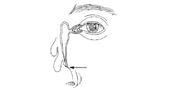
A. Valve of Krause
B. Valve of Hasner
C. Valve of Reitzius
D. None of the above
Show Answer
|
Correct Answer » B Explanation |
 |
Ans:B. Valve of Hasner
Nasolacrimal drainage system
- It serves as a conduit for tear flow from the external eye to the nasal cavity.
- It consists of the puncta, canaliculi, lacrimal sac, and nasolacrimal duct .
- Puncta are openings 0.3 mm in diameter located on the medial aspect of the upper and lower eyelid margins.
- The inferior punctum is approximately 0.5 mm lateral to the superior punctum, with distances to the medial canthus of 6.5 mm and 6.0 mm, respectively. Tears within the medial canthal area enter the puncta to pass into the canaliculi.
- Canaliculi have an initial vertical segment, measuring 2 mm, followed by an 8-mm horizontal segment .The angle between the vertical and horizontal segments is approximately 90 degrees.
- In most individuals, the horizontal portion of the canaliculi converges to form the common canaliculus. Canaliculi pierce the lacrimal fascia before entering the lacrimal sac.
- The lacrimal sac sits within the lacrimal fossa, which is bound anteriorly by the frontal process of the maxillary bone (anterior lacrimal crest) and posteriorly by the lacrimal bone (posterior lacrimal crest).
- The nasolacrimal duct consists of a 12-mm superior intraosseous portion and a 5-mm inferior membranous portion. It opens into the inferior meatus under the inferior nasal turbinate. The opening of the nasolacrimal duct into the inferior nasal meatus of the nasal cavity is partially covered by a mucosal fold (valve of Hasner or plica lacrimalis).
