Degradation of Purine Nucleotides
Uric acid is formed in humans in
A |
Liver |
|
B |
Kidney |
|
C |
GIT mucosa |
|
D |
Joints |
Uric acid is formed in humans in
A |
Liver |
|
B |
Kidney |
|
C |
GIT mucosa |
|
D |
Joints |
GIT mucosa
Most of the dietary purines are converted to uric acid in lhe intestinal’mucosal cell only. Intestinal bacterialJlora is intolved in degradation of the rest of dietary purines that remain unabsorbed.
A patient with increased Hypoxanthine and Xanthine in blood with hypouricemia which enzyme is deficient?
A |
HGPRTase |
|
B |
Xanthine oxidase |
|
C |
Adenosine deaminase |
|
D |
APRtase |
A patient with increased Hypoxanthine and Xanthine in blood with hypouricemia which enzyme is deficient?
A |
HGPRTase |
|
B |
Xanthine oxidase |
|
C |
Adenosine deaminase |
|
D |
APRtase |
- Hypouricemia- Hypouricemia and increased excretion of hypoxanthine and xanthine are associated with xanthine oxidase deficiency.
- Lesch-Nyhan Syndrome- The Lesch-Nyhan syndrome, an overproduction hyperuricemia characterized by frequent episodes of uric acid lithiasis and a bizarre syndrome of selfmutilation reflects a defect in hypoxanthine-guanine phosphoribosyltransferase, an enzyme of purine salvage.
- Adenosine Deaminase Deficiency- Adenosine deaminase deficiency is associated with an immunodeficiency disease in which both thymus-derived lymphocytes (T cells) and bone marrow-derived lymphocytes (B cells) are sparse and dysfunctional. Patients suffer from severe immunodeficiency.
- Purine Nucleoside Phosphorylase Deficiency- Purine nucleoside phosphorylase deficiency is associated with a severe deficiency of T cells but apparently normal B cell function
Degradation of Purine Nucleotides
CATABOLISM OF PURINE NUCLEOTIDES
- Human catabolises purine to uric acid.
- In higher primates, Allantoin by enzyme uricase is the end product.
- Adenine nucleotides catabolism- liver, heart muscle, Skeletal muscle, GIT mucosa.
- Guanine nucleotides catabolism- liver, spleen, kidney, pancreas, GIT mucosa.
- First metabolic product of purines is Xanthine.

Exam Important
- Human catabolise purines (adenine and guanine) to uric acid.
- First metabolic product of purines is xanthine.
- In non primate mammals, uric acid is converted to water soluble allantion by enzyme uricase.
- The end product of purine catablism is uric acid, which is excreted.
Degradation of Pyrimidine Nucleotides
What is the end product of catabolism of pyrimidine?
A |
NH3 |
|
B |
CO2 & H2O |
|
C |
Both |
|
D |
None |
What is the end product of catabolism of pyrimidine?
A |
NH3 |
|
B |
CO2 & H2O |
|
C |
Both |
|
D |
None |
The end products of pyrimidine catabolism is CO2 and H2O.
Which is the product excreted unchanged in catabolism of pyrimidine?
A |
Uric acid |
|
B |
NH3 |
|
C |
Pseudouridine |
|
D |
Beta alanine |
Which is the product excreted unchanged in catabolism of pyrimidine?
A |
Uric acid |
|
B |
NH3 |
|
C |
Pseudouridine |
|
D |
Beta alanine |
Pseudouridine is excreted unchanged as it cannot be catabolized in human.
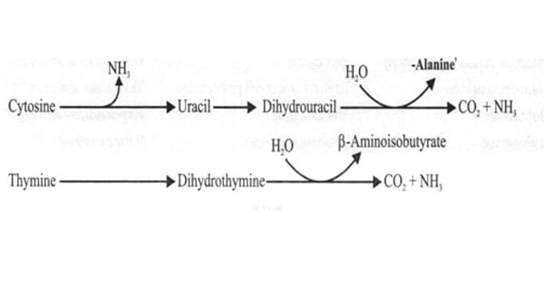
Degradation of Pyrimidine Nucleotides
CATABOLISM OF PYRIMIDINE NUCLEOTIDES
- Cytosine and Uracil to Beta Alanine — CO2, NH3
- Thymine and β- aminoisobutyrate — CO2, NH3
- The end products are highly water soluble.

Exam Important
- The end products of pyrimidine catabolism are highly water soluble. E.g. CO2, NH3, β- aminoisobutyrate, beta alanine.
- Pseudouridine is excreted unchanged as it cannot be catabolized in human.
- Humans have no enzyme that can catabolise pseudouridine derived from degradation of t-RNA.
- There is no energy generated in pyrimidine catabolism.
- The end products of pyrimidine catabolism is CO2 and H2O.
Steps Of Protein Synthesis (Translation)
A | Ribosomal RNA | |
B | Messenger RNA | |
C | Signal recognition particle | |
D | Peptidyl transferase |
A | Ribosomal RNA | |
B | Messenger RNA | |
C | Signal recognition particle | |
D | Peptidyl transferase |
Signal recognition particles (SRPs) recognize the signal sequence on the N-terminal end of proteins destined for the lumen of the endoplasmic reticulum (ER). SRP binding arrests translation and an SRP receptor facilitates import of the SRP, ribosome, and nascent protein into the ER lumen. A signal peptidase removes the signal sequence from the protein, which may remain in the membrane or be routed for secretion.
Common to both eukaryotic and prokaryotic protein synthesis is the requirement for ATP to activate amino acids. The activated aminoacyltRNAs then interact with ribosomes carrying mRNA. Peptidyl transferase catalyzes the formation of peptide bonds between the free amino group of activated aminoacyl-tRNA on the A site of the ribosome and the esterified carboxyl group of the peptidyl-rRNA on the P site; the liberated rRNA remains on the P site.
GTP is required by which of the following steps in protein synthesis?
A |
Aminoacyl—tRNA synthetase activation of amino acids |
|
B |
Attachment of mRNA to ribosomes |
|
C |
Attachment of ribosomes to endoplasmic reticulum |
|
D |
Translocation of tRNA—nascent protein complex from A toP sites |
GTP is required by which of the following steps in protein synthesis?
A |
Aminoacyl—tRNA synthetase activation of amino acids |
|
B |
Attachment of mRNA to ribosomes |
|
C |
Attachment of ribosomes to endoplasmic reticulum |
|
D |
Translocation of tRNA—nascent protein complex from A toP sites |
he entry of the aminoacyl-tRNA into the A site results in the hydrolysis of one GTP to GDP.
Translocation of the newly formed peptidyl-tRNA in the A site into the P site by EF 2 similarly results in hydrolysis of GTP to GDP and phosphate.
Thus, the energy requirements for the formation of one peptide bond include the equivalent of the hydrolysis of two ATP molecules to ADP and of two GTP molecules to GDP, or the hydrolysis of four high-energy phosphate bonds.
Protein synthesis in eukaryotes is different from prokaryotes. Which of the following is NOT TRUE about steps in initiation of eukaryotic protein synthesis?
A |
Dissociation of the ribosome into its 40S and 60S subunits |
|
B |
Binding of a ternary complex to the 40S ribosome |
|
C |
Binding of mRNA to the 60S preinitiation complex to form the 43S initiation complex |
|
D |
Combination of the 48S initiation complex with the 60S ribosomal subunit |
Initiation of protein synthesis can be divided into four steps:
- Dissociation of the ribosome into its 40S and 60S subunits;
- Binding of a ternary complex consisting of the initiator methionyl-tRNA, (met-tRNAi), GTP, and eIF-2 to the 40S ribosome to form the 43S preinitiation complex;
- Binding of mRNA to the 40S preinitiation complex to form the 48S initiation complex; and
- Combination of the 48S initiation complex with the 60S ribosomal subunit to form the 80S initiation complex.
Chain initiation in protein synthesis is by‑
A |
AUG |
|
B |
GLA |
|
C |
UGA |
|
D |
UAG |
Chain initiation in protein synthesis is by‑
A |
AUG |
|
B |
GLA |
|
C |
UGA |
|
D |
UAG |
A i.e. AUG
The cellular component for protein synthesis is :
A |
Smooth endoplasmic reticulum |
|
B |
Rough endoplasmic reticulum |
|
C |
Ribosomes |
|
D |
Mitochondria |
The cellular component for protein synthesis is :
A |
Smooth endoplasmic reticulum |
|
B |
Rough endoplasmic reticulum |
|
C |
Ribosomes |
|
D |
Mitochondria |
C i.e. Ribosomes
Termination process of protein synthesis is performed by:
A |
Releasing factor |
|
B |
Stop codon |
|
C |
UAA codon |
|
D |
All |
Termination process of protein synthesis is performed by:
A |
Releasing factor |
|
B |
Stop codon |
|
C |
UAA codon |
|
D |
All |
A, B, C i.e. Releasing factor, Stop codon, UAA codon
Which of the following is not required for protein synthesis of eukaryotes:
A |
RNA polymerase |
|
B |
Ribosomes |
|
C |
Peptidyl transferase |
|
D |
Amino acyl tRNA synthetase |
Which of the following is not required for protein synthesis of eukaryotes:
A |
RNA polymerase |
|
B |
Ribosomes |
|
C |
Peptidyl transferase |
|
D |
Amino acyl tRNA synthetase |
Ans. a. RNA polymerase (Ref Harper 28/e p362) RNA polymerase enzyme is involved in transcription process, not in translation.
The a-amino group of the new aminoacyl-tRNA in the A site carries out a nucleophilic attach on the esterified carboxyl group of the peptidyl-tRNA occupying the P site (peptidyl or polypeptide site).
Peptidyl transferase: Catalyses two reactions, peptide bond formation between amino acids and together with release factor, peptide release.
False about eukaryotic protein synthesis is:
A |
N formyl Met is the first-RNA to come into action |
|
B |
mRNA read from 5′ to 3′ |
|
C |
Eft shift between GDP to GTP |
|
D |
Capping helps in attachment of mRNA to 40 S ribosome |
False about eukaryotic protein synthesis is:
A |
N formyl Met is the first-RNA to come into action |
|
B |
mRNA read from 5′ to 3′ |
|
C |
Eft shift between GDP to GTP |
|
D |
Capping helps in attachment of mRNA to 40 S ribosome |
A i.e. N formyl Met is the first – RNA to come into action
Protein synthesis occurs in:
September 2012
A |
Smooth endoplasmic reticulum |
|
B |
Rough endoplasmic reticulum |
|
C |
Golgi bodies |
|
D |
Nucleus |
Protein synthesis occurs in:
September 2012
A |
Smooth endoplasmic reticulum |
|
B |
Rough endoplasmic reticulum |
|
C |
Golgi bodies |
|
D |
Nucleus |
Ans. B i.e. Rough endoplasmic reticulum
Rough ER
- The surface of the rough endoplasmic reticulum (often abbreviated RER) is studded with protein-manufacturing ribosomes giving it a “rough” appearance (hence its name).
STEPS OF PROTEIN SYNTHESIS (Translation)
TRANSLATION
- Translation is the process in which the genetic information stored in DNA is passed on to mRNA where it is translated into proteins.
- Translation occurs in ribosomes.
- mRNA is translated from its 5’ end to its 3’- end (51 à 31 )
- 4 letter language information from nucleic acids to 20 letter language proteins.
STEPS OF PROTEIN SYNTHESIS-
- Activation of Amino acid
- Initiation
- Elongation
- Termination
1. Activation of amino acid (charging of tRNA)
- Activation of amino acids takes place cytosol.
- Each of the 20 amino acids covalently attached to the respective tRNA, by the ATP as two high energy phosphate bond catalyzed by aminoacyl tRNA synthase (AAS) so called as charging tRNA.
- AAS is identified by DHU arm and is considered as proofreading mechanism of translation.

- Aminoacyl tRNA synthase – are specific for particular amino acids and tRNA.
- They are responsible for high fidelity of translation of genetic message.
- Implements genetic code by acting as molecular dictionaries.
- 2 ATPs are required for this reaction.
2. Initiation- is a multi process stage.
- It is facilitated by accessory proteins called Initiation factors (IF) and for eukaryotic initiation factor (eIF).
a) Ribosomal dissociation–
- Two initiation factors(eIF3 & eIF-1A) binds to 40S subunit of eukaryotic ribosome (80S).
- 80S ribosomes disassociates into 40S and 60S subunits.
b) Formation of 43S preinitiation complex–
- A ternary complex containing met- tRNA1 and eIF-2(controlling factor in eukaryotes) bound to GTP attaches to 40S subunit to form 43S preinitiation complex.
- AUG serves as initiation codon for protein synthesis and codes for methionine
c)Formation of 48S Initiation complex-
- The binding of mRNA to 43S preinitiation complex results in the formation 48S initiation complex is facilitated by 7- methyguanylate cap at 51 –end of mRNA.
- In Eukaryotes, Kozak consensus sequence surrounds AUG and determines the initiating codon of mRNA.
- In Prokaryotes, a sequence of nucleotide bases on mRNA called as Shine- Dalgarno sequence (SD sequence). It is located -6 to -10bp from AUG codon. (purine rich)
d) Formation of 80S initiation complex-
- 48S initiation complex + 60S subunit = 80S initiation complex
- 3 sites on 80S Ribosome- A site, P site, E site.

3. Elongation-
- Catalyzed by proteins called as elongation fators.
- Has 4 steps-
a) Binding of aminoacyl tRNA to the A-site-
- Elongation factor EF-1 helps in binding of tRNA.
b) Peptide bond formation-
- The methionine of tRNA of P- site is transferred to the new amino acid on tRNA of A-site to form peptide bond catalyzed by peptidyl transferase (a ribozyme).
c) Translocation-
- It requires elongation factor eEF2 (translocase) and hydrolysis of GTP.
4. Termination-
- Stop codon is in the A site now.
- In eukaryotes, one single releasing factor, eRF.
- In prokaryotes, 3 releasing factors- RF- 1, RF-2, RF-3.
Exam Important
- Translation occurs in ribosomes.
- mRNA is translated from its 5’ end to its 3’- end (51 à 31 )
- AAS is identified by DHU arm and is considered as proofreading mechanism of translation.
- Aminoacyl synthase implements genetic code by acting as molecular dictionaries.
- Two initiation factors(eIF3 & eIF-1A) binds to 40S subunit of eukaryotic ribosome (80S).
- AUG serves as initiation codon for protein synthesis and codes for methionine
- Shine- Dalgarno sequence located -6 to -10bp from AUG codon. (purine rich).
- 3 sites on 80S Ribosome-
- A site- new aminoacyl tRNA binds
- P site- growing peptidyl chain present
- E site- deacylated tRNA present.
- 2 ATPs are required for activation of amino acid.
- There is no tRNA for hydroxyproline and hydroxylysine.
Acute myeloid leukemia (AML)
Non-specific esterase is positive in all the categories of Acute Myeloid Leukemia, EXCEPT:
A |
M3 |
|
B |
M4 |
|
C |
M5 |
|
D |
M6 |
Non-specific esterase is positive in all the categories of Acute Myeloid Leukemia, EXCEPT:
A |
M3 |
|
B |
M4 |
|
C |
M5 |
|
D |
M6 |
Non specific esterase is negative in M3 type of Acute myeloid leukemia (AML).
Alpha naphthyl acetate esterase (ANAE), Alpha naphthyl butyrate esterase (ANBE) and Alpha naphthyl AS esterase (NASA) are the non specific esterase reactions that are positive in M4 (acute myelomonocytic leukemia) and M5 (acute monocytic/monoblastic leukemia) type of AML.
Their presence is variable in M6 (acute erythroid leukemia).
Ref: Wintrobe’s Clinical Hematology, 10th Ed, Page 2221; Textbook of pathology, B N Datta, 2nd Edition, Page 1118 & 1119.
A 32 year old male is diagnosed of having acute myeloid leukemia. His total WBC count was less than normal initially. Which of the following factor has a bad prognosis in AML?
A |
Monosomies of chromosomes |
|
B |
Young age |
|
C |
Patients with t(15;17) |
|
D |
Low WBC count |
A 32 year old male is diagnosed of having acute myeloid leukemia. His total WBC count was less than normal initially. Which of the following factor has a bad prognosis in AML?
A |
Monosomies of chromosomes |
|
B |
Young age |
|
C |
Patients with t(15;17) |
|
D |
Low WBC count |
Prognostic factors of acute myeloid leukemia (AML):
- Advancing age is associated with a poorer prognosis, in part because of its influence on the patient’s ability to survive induction therapy.
- The leukemic cells in elderly patients more commonly express the multidrug resistance 1 (MDR1) efflux pump that conveys resistance to natural product–derived agents such as the anthracyclines.
- Patients with t(15;17) have a very good prognosis, and those with t(8;21) and inv(16) a good prognosis, while those with no cytogenetic abnormality have a moderately favorable outcome.
- Patients with a complex karyotype, t(6;9), inv(3), or -7 have a very poor prognosis.
- Among patients with hyperleukocytosis (>100,000/L), early central nervous system bleeding and pulmonary leukostasis contribute to poor outcome with initial therapy.
- Karyotypes include monosomy chromosome 5 or chromosome 7 have 78% of relapse rate.
All of the following genetic syndromes are associated with Acute Myeloid Leukemia, except:
A |
Down’s Syndrome |
|
B |
Klinefelter’s Syndrome |
|
C |
Patau Syndrome |
|
D |
Turner’s Syndrome |
All of the following genetic syndromes are associated with Acute Myeloid Leukemia, except:
A |
Down’s Syndrome |
|
B |
Klinefelter’s Syndrome |
|
C |
Patau Syndrome |
|
D |
Turner’s Syndrome |
Of all the options given Turner’s syndrome is not found to be associated with increased incidence of acute myeloid leukemia (AML). Down’s syndrome, Klinefelter’s Syndrome, Patau Syndrome are associated with AML.
Ref: Harrison’s Principles of Internal Medicine16th Edition, Page 631; Excellent Care for Cancer Survivors: A Guide to Fully Meet Their Needs By Kenneth Miller – Pg 327; Childhood Leukemia: A Practical Handbook, By Gregory H. Reaman – Pg 11
All of the following are poor prognostic factors in a case of acute myeloid leukemias, except:
A |
Age more than 60 years |
|
B |
Presence of t(8:21) |
|
C |
Secondary leukemias |
|
D |
Leucocyte count more than 1,00,000/microl |
All of the following are poor prognostic factors in a case of acute myeloid leukemias, except:
A |
Age more than 60 years |
|
B |
Presence of t(8:21) |
|
C |
Secondary leukemias |
|
D |
Leucocyte count more than 1,00,000/microl |
Patients t(8;21) and inv(16) has a good prognosis, approximately 55% of these patients are cured. t(15:17) is associated with a very good prognosis, while those without any cytogenetic abnormality have a moderately favorable outcome.
Which of the following is a poor prognostic factor in Acute Myeloid Leukemia (AML)
A |
Monosomy |
|
B |
Deletion of X or Y chromosome |
|
C |
t (8; 21) translocation |
|
D |
Nucleophosphin mutation |
Which of the following is a poor prognostic factor in Acute Myeloid Leukemia (AML)
A |
Monosomy |
|
B |
Deletion of X or Y chromosome |
|
C |
t (8; 21) translocation |
|
D |
Nucleophosphin mutation |
Answer is A (Monosomy)
Monosomy is consistently associated with an unfavorable or poor prognosis.
Monosomy is associated with a poor prognosis
Monosomy especially those involving chromosome 7 (monosomy 7) and chromosome 5 (monosomy 5) are consistently associated with poor prognosis in both adults and children with AML
Deletion of X or Y chromosome is associated with a favorable / intermediate prognosis
‘Monosomy of the X chromosome in a female patient (loss of the Y chromosome) is the most common whole chromosome loss identified in pediatric patients with AML. This numeric abnormality is usually associated with t(8; 21) translocation and AML M2 which carry a good prognosis’ – ‘Childhood Leukemias’ by Puri 2″d/253
`Loss of Y and X chromosomes are most frequently observed in patients with t(8; 21) which carries a favourable prognosis’ – ‘Blood: Principles and Practice of Hematology’ 2″d/108
Nucleophosphin mutation is associated with a favorable prognosis |
||
|
||
Factor |
Favourable |
Unfavourable |
Nucleophosphin mutation |
Present |
Absent |
t (8; 21) translocation is associated with a favorable prognosis |
Factor |
Favourable |
Unfavourable |
Cytogenetics |
t(15;17), 1(8;21), inv(16) |
-7, del(7q), -5, del(5q), 3q21 and 3q26 abnormalities, complex karyotypes |
Prognostic Feature in Acute Myeloid Leukemia:
Factor Favourable Unfavourable |
||
Clinical |
||
Age |
<45 yr |
<2yr, >60yr |
ECOG performance status |
0-1 |
> I |
Leukemia |
De novo |
Antecedent hematologic disorder, myelodysplasia, myeloproliferative disorder |
Infection |
Absent |
Present |
Prior chemotherapy |
No |
Yes |
Leukocytosis |
<25,000/mm3 |
> 100,000/mm2 |
Serum LDH |
Normal |
Elevated |
Extramedullary disease |
Absent |
Present |
CNS disease |
Absent |
Present |
Cytoreduction |
Rapid |
Delayed |
Morphology |
||
Auer rods |
Present |
Absent |
Eosinophils |
Present |
Absent |
Megaloblastic erythroids |
Absent |
Present |
Dysplastic megakaryocytes |
Absent |
Present |
FAB type |
M2, M3, M4 |
MO, M6, M7 |
Surface/enzyme markers |
||
Myeloid |
CD34-, CDI4-, CD13- |
CD34+ |
HLA-DR |
Negative |
Positive |
TdT |
Absent |
Present |
Lymphoid |
Cd2+ |
CD7+, CD56+ Biphenotypic (2 or more lymphoid markers) Present |
MDR-1 |
Absent |
|
Cytogenetics |
||
Cytogenetics |
1(15;17), 1(8;21), inv(16) |
-7, del(7q), -5, del(5q), 3q21 and 3q26 abnormalities, complex karyotypes |
Molecular markers |
||
Fms-related tyrosine kinase-3 mutation |
Absent |
Present |
Ecotropic viral integration site 1 expression |
Absent |
Present |
Mixed-lineage leukemia partial tandem duplication |
Absent |
Present |
Nucleophosphin mutation |
Present |
Absent |
CCAAT/enhancer-binding protein- a mutation |
Present |
Absent |
Brain and acute leukemia cytoplasmic gene expression |
Absent |
Present |
Vascular endothelial growth factor expression |
Absent |
Present |
In acute myeloid leukemia, Auer rods are numerous in:
September 2009
A |
M2 |
|
B |
M3 |
|
C |
M4 |
|
D |
M5 |
In acute myeloid leukemia, Auer rods are numerous in:
September 2009
A |
M2 |
|
B |
M3 |
|
C |
M4 |
|
D |
M5 |
Ans. B: M3
The diagnosis of AML is based on the presence of at least 20% myeloid blasts in the bone marrow. Myeloblast have delicate nuclear chromatin, two to four nucleoli, and more voluminous cytoplasm than lymphoblasts.
The cytoplasm often contains fine, peroxidase-positive azurophilic granules.
Auer rods, distinctive needle like azurophilic granules, are present in many cases; they are particularly numerous in AML with the t(15;17) (acute promyelocytic leukaemia-M3).
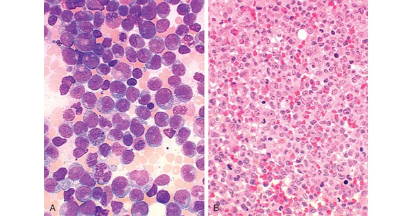
Acute myeloid leukemia (AML)
ACUTE MYELOID LEUKEMIA (AML)
- AML is a heterogenous disease characterised by infiltration of malignant myeloid cells into blood, bone marrow.
- AML is due to inhibition of maturation of myeloid stem cells due to mutations.
- Seen in mainly in adults (50 years).
- Chromosomal mutations in AML are
- translocation t (8: 21) & t (15: 17)
- Inversion 16 or t (16: 16)

Etiology-
- Hereditary – Down syndrome, Klinefelter’s Syndrome, Patau Syndrome.
- Radiation
- Chemical- smoking
Pathogenesis-
- t (8: 21) disrupt the RUNXL gene & Inv (16) disrupts the CDF1β gene both have good prognosis.
- t (15: 17) (acute promyelocytic leukaemia-M3) have good prognosis.
- Gene mutation encoding components of cohesion complex.
- Most common congenital AML (in infants) are AML M5 (acute monocytic leukemia)
- Most common AML in children is AML M7 (acute megakaryoblastic anaemia)
- Most common translocation- MLL gene rearrangements on chromosome 11q.
- Monosomy is associated with a poor prognosis.
Clinical features-
1. Due to bone marrow failure-
- Anaemia
- Bruises, petechiae, bleeding from gum
- Infection
- Fever
2. Due to organ infiltration-
- Pain & tenderness of bones
- Lymphadenopathy, enlargement of tonsils
- Splenomegaly
- Hepatomegaly
- Gum hypertrophy
- Chloroma

Investigations-
1. Blood picture-
- Anemia
- Thrombocytopenia
- WBC increased
2. Bone marrow examination
- Cellularity- marrow is hypercellular but blood tap or dry tap is seen.
- Leukemic cells- Blast cell count >20% (WHO)
- Dyserythropoiesis, megaloblastic features & ring sideroblasts are common.
- Megakaryocytes.
3. Cytochemistry-
- Myeloperoxidase- Positive in immature myeloid cells containing granules & Auer rods (most definitive sign of myeloid differentiation)
- Cluster of Auer rods called as Faggot.
- Auer rods, distinctive needle like azurophilic granules,they are particularly numerous in AML with the t (15: 17) (acute promyelocytic leukaemia-M3).
- Non specific esterase (NSE)- positive in monocytic series (M3, M4 & M5)
- Investigation of choice is flow cytometry.


Treatment-
- Blood transfusion & platelet transfusion.
- Cytotoxic drug therapy- most effective treatment of AML is cytosine, arabinoside, anthracyclines.
- Promyelocytic leukemia (M3)- tretinoin orally
- Bone marrow transplantation.
Exam Important
- AML is due to inhibition of maturation of myeloid stem cells due to mutations.
- Seen in mainly in adults (50 years).
- Chromosomal mutations in AML are
- translocation t (8: 21) & t (15: 17)
- Inversion 16 or t (16: 16)
Pathogenesis-
- t (8: 21) disrupt the RUNXL gene
- Inv (16) disrupts the CDF1β gene.
- Gene mutation encoding components of cohesion complex.
- Most common congenital AML (in infants) are AML M5 (acute monocytic leukemia)
- Most common AML in children is AML M7 (acute megakaryoblastic anaemia)
- Most common translocation- MLL gene rearrangements on chromosome 11q.
Investigations-
- Leukemic cells- Blast cell count >20% (WHO)
- Dyserythropoiesis, megaloblastic features & ring sideroblasts are common
- Myeloperoxidase- Positive in immature myeloid cells containing granules & Auer rods (most definitive sign of myeloid differentiation)
- Cluster of Auer rods called as Faggot.
- Non specific esterase (NSE)- positive in monocytic series (M3, M4 & M5)
- Investigation of choice is flow cytometry
Treatment-
- Cytotoxic drug therapy- most effective treatment of AML is cytosine, arabinoside, anthracyclines.
Sjogren syndrome
Biopsy of the parotid gland in a patient with Sjogren’s syndrome shows –
A |
Neutrophils |
|
B |
Lymphocytes |
|
C |
Eosinophi Is |
|
D |
Basophils |
Biopsy of the parotid gland in a patient with Sjogren’s syndrome shows –
A |
Neutrophils |
|
B |
Lymphocytes |
|
C |
Eosinophi Is |
|
D |
Basophils |
Ans. is ‘b’ i.e., Lymphocytes
o The earliest histological finding in both the major and minor salivary glands is periductal and perivascular lymphocytic infilteration which eventually becomes extensive
Sjogren’s syndrome refers to disease of ‑
A |
Parotid glands |
|
B |
Thyroid disease |
|
C |
Parathyroid glands |
|
D |
Multiple endocrine neoplasia |
Sjogren’s syndrome refers to disease of ‑
A |
Parotid glands |
|
B |
Thyroid disease |
|
C |
Parathyroid glands |
|
D |
Multiple endocrine neoplasia |
Ans. is ‘a’ i.e., Parotid glands
All of the following are true about Primary Sjogren’s syndrome, except:
A |
May be seen in children |
|
B |
Sensation of sand or gravel in eyes |
|
C |
Associated with rheumatoid arthritis |
|
D |
Salivary gland enlargement |
All of the following are true about Primary Sjogren’s syndrome, except:
A |
May be seen in children |
|
B |
Sensation of sand or gravel in eyes |
|
C |
Associated with rheumatoid arthritis |
|
D |
Salivary gland enlargement |
Answer is C (Associated with Rheumatoid Arthritis):
Kelly’s Rheumatoid arthritis is associated with Secondary Sjogren’s Syndrome and not Primary Sjogren’s Syndrome.
Primary Sjogren’s Syndrome |
No Connective Tissue /Chronic inflammatory disorder |
Secondary Sjogren’s Syndrome |
Underlying Connective Tissue / Chronic inflammatory disorder |
Keratoconjunctivitis Sicca (Sensation of sand or gravel in eyes) and salivary gland enlargement are typical symptoms of Sjogren’s syndrome (Both Primary and Secondary). Primary Sjogren’s syndrome typically affects women in their middle age (Female to male ratio = 9:1) but it may occur at any age including childhood. Primary Sjogren’s syndrome
has been reported infrequently in children with onset as early as 5 years of age. The presence of symptoms and signs of Sjogren’s ‘s syndrome (Dry eyes; Dry mouth; Salivary Gland Enlargement) in the setting of another connective tissue disease or chronic inflammatory pathology like Rheumatoid Arthritis, SLE, Systemic Sclerosis by definition is termed as Secondary Sjogren’s Syndrome.
All of the following are features of Sjogren’s syndrome except:
A |
It is an autoimmune chronic inflammatory disease |
|
B |
Typically occurs in women after the menopause |
|
C |
In primary Sjogren’s syndrome, keratoconjunctivitis sicca is associated with rheumatoid arthritis |
|
D |
In secondary Sjogren’s syndrome, dry eye and/ or xerostomia (dry mouth) is associated with rheumatoid arthritis |
Ans. In primary Sjogren’s syndrome, keratoconjunctivitis sicca is associated with rheumatoid arthritis
Regarding Sjogren’s syndrome, all are true except:
September 2010
A |
Keratoconjuctivitis sicca |
|
B |
Rheumatoid arthritis |
|
C |
Epiphora |
|
D |
Autoimmune in nature |
Regarding Sjogren’s syndrome, all are true except:
September 2010
A |
Keratoconjuctivitis sicca |
|
B |
Rheumatoid arthritis |
|
C |
Epiphora |
|
D |
Autoimmune in nature |
Ans. C: Epiphora
SjOgren’s syndrome (also known as “Mikulicz disease” and “Sicca syndrome”, is a systemic autoimmune disease in which immune cells attack and destroy the exocrine glands that produce tears and saliva
SjOgren’s syndrome can exist as a disorder in its own right (Primary Sjogren’s syndrome) or it may develop years after the onset of an associated rheumatic disorder such as rheumatoid arthritis, systemic lupus erythematosus, scleroderma, primary biliary cirrhosis etc. (Secondary SjOgren’s syndrome)
True regarding Sjogren’s syndrome are all of the following except:
September 2009
A |
Autoimmune condition |
|
B |
Males are commonly affected |
|
C |
Progressive destruction of lacrimal and salivary gland |
|
D |
No single laboratory investigation is pathognomic |
True regarding Sjogren’s syndrome are all of the following except:
September 2009
A |
Autoimmune condition |
|
B |
Males are commonly affected |
|
C |
Progressive destruction of lacrimal and salivary gland |
|
D |
No single laboratory investigation is pathognomic |
Ans. B: Males are commonly affected
SjOgren’s Syndrome (SS) is a systemic autoimmune disease characterised by lymphocytic infiltration, acinar cell destruction and proliferation of duct epithelium in all salivary and larimal gland tissue.
Extra glandular involvement of muscles, blood vessels, lungs, kidneys may also occur.
Females are affected more than the males
It may be primary and secondary (when associated with other connective tissue diseases). There is a risk of progression to lymphoid malignancy.
Dry mouth, dry eyes and arthritis/arthralgia are the 3 common presenting features.
Of the extraarticular manifestations, Raynaud’s phenomena is the most common skin manifestation seen in 35% of patients. Vasculitis has been reported in 5% of patients with Sjogren’s syndrome. This includes small vessel leucocytoclastic vasculitis and medium vessel necrotising vasculitis.
Distal RTA may be silent or lead to renal stones, nephrocalcinosis and compromised renal function. Hypergammaglobulinemia may be due to polyclonal activation of B cells.
The diagnosis is base on the history as no single laboratory investigation is pathognomic of either primary or secondary Sjogren’s syndrome
Development of Lymphoma in Sjogren’s syndrome is suggested by all of the following except
A |
Persistent parotid gland enlargement |
|
B |
Cyoglobilinemia |
|
C |
Leukopenia |
|
D |
High C4 compement levels |
Development of Lymphoma in Sjogren’s syndrome is suggested by all of the following except
A |
Persistent parotid gland enlargement |
|
B |
Cyoglobilinemia |
|
C |
Leukopenia |
|
D |
High C4 compement levels |
Ans. is ‘d’ i.e., High C4 complement levels
- Lymphoa is a well-known complication of Sjogren’s syndrome Most lymphomas are extra-nodal, low grade marginal B cell lymphomas.
- Development of Lymphoma in Sjogren’s syndrome is suggested by low C4 complement levels.
Lymphoma in Sjogren’s syndrome
The development ofLymphomas in patients with Sjogren syndrome is suggested by : –
- Persistent parotid gland enlargement
- Purpura
- Leukopenia
- Cryoglobulinemia
- Low C4 complement levels
SJOGREN’S SYNDROME
SJOGREN’S SYNDROME
- Sjogren’s syndrome is an autoimmune disorder associated with parotid glands.
- It is characterised by lymphocytic infiltration of salivary and lacrimal gland.
- It affects women more (40- 60 years)
- Associated with HLA- B8 & DR3.
- Associated with RA, SLE, and primary biliary cirrhosis.
Clinical Features-
- Dry eyes ( keratoconjuctivitis sicca)
- Xerostomia
- Vaginal dryness
- Raynaud’s phenomenon
- Lymphoma
- Splenomegaly

The development ofLymphomas in patients with Sjogren syndrome is suggested by : –
- Persistent parotid gland enlargement
- Purpura
- Leukopenia
- Cryoglobulinemia
- Low C4 complement levels
Investigations-
- Schirmer test- for flow of tear
- ESR elevated
- Biopsy- The earliest histological finding in both the major and minor salivary glands is periductal and perivascular lymphocytic infilteration which eventually becomes extensive
- Autoantibodies- RF, ANA, anti- Ro (SS-A), anti- La (SS- B)
Treatment-
- Lubricants for dry eyes.
- Xerostomia- saliva forming lubricants
- Corticosteroids for extraglandular & musculoskeletal deformities.
Exam Important
- Sjogren’s syndrome is an autoimmune disorder associated with parotid glands.
- It is characterised by lymphocytic infiltration of salivary and lacrimal gland.
- It affects women more (40- 60 years)
- Associated with HLA- B8 & DR3.
- Associated with RA, SLE, and primary biliary cirrhosis.
Clinical Features-
- Dry eyes ( keratoconjuctivitis sicca)
- Xerostomia
- Vaginal dryness
- Raynaud’s phenomenon
- Lymphoma
- Splenomegaly
The development ofLymphomas in patients with Sjogren syndrome is suggested by : –
- Persistent parotid gland enlargement
- Purpura
- Leukopenia
- Cryoglobulinemia
- Low C4 complement levels
Investigations-
- Autoantibodies- RF, ANA, anti- Ro (SS-A), anti- La (SS- B)